神经退行性病
-
Figure 1|Formation of exosomes.
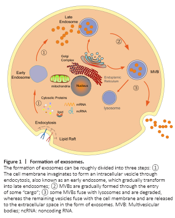
Exosomes refer to a special subtype of nanoscale EVs that were first discovered during the culturing and differentiation of sheep reticulocytes in the late 1980s (Johnstone et al., 1987) and were first detected in the blood of healthy people in 2005 (Ren et al., 2011). Exosomes are secreted from the cytoplasmic membrane into the extracellular environment due to the budding of the cytoplasmic membrane. The formation of exosomes can be roughly divided into three steps (Simons and Raposo, 2009; Bellingham et al., 2012; Rufino-Ramos et al., 2017; Hessvik and Llorente, 2018; Ortega et al., 2020), as shown in Figure 1.
Figure 2|Exosomes, α-synuclein (α-syn), and glial cells involved in the mechanisms underlying neuroinflammation and neurodegenerat
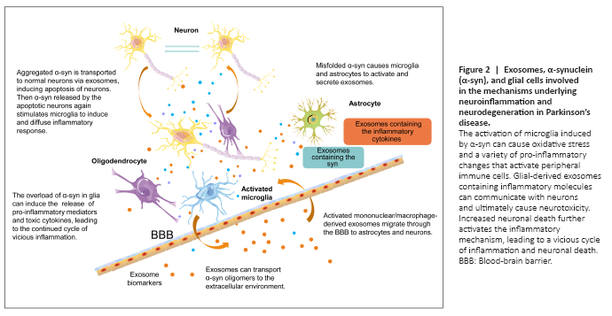
Neurons and other types of cells, including astrocytes, can internalize α-synuclein released by pathological neurons, resulting in the formation of pathological inclusion bodies and subsequent inflammation in the recipient cells (Lee et al., 2010). Under healthy conditions, astrocytes express α-synuclein at low levels or not at all (Lee et al., 2010). Misfolded α-synuclein causes microglia to activate and release toxic cytokine factors, which can metabolize and cleave natural α-synuclein, inducing misfolding and aggregation and resulting in the formation of pathological inclusion bodies (Guo et al., 2020). Misfolded synaptic proteins are released from cells and can be ingested by microglia, which causes them to be activated by the release of toxic cytokines, continuing a vicious circle. The release of various toxic cytokines by microglia can lead to the misfolding of α-synuclein in nearby cells, promoting the selective transmission and reproduction of synuclein pathology in vulnerable cell populations (Olanow et al., 2019) (Figure 2). In general, exosomes may be involved in various stages of the inflammatory process, including neuron-to-neuron communication, neuron-to-glia communication, and glia-to-glia communication. Exosomes provide an environment for α-syn aggregation and may promote the aggregation of α-syn oligomers. α-Syn induces the activation of microglia and astrocytes, and activated microglia, astrocytes, and T cells can interact to increase the release of humoral and pro-inflammatory cytokines, aggravate neuroinflammation and ROS production, and promote neuronal death (Yu et al., 2020) in a vicious cycle that exacerbates PD disease progression (Figure 2).