视神经损伤
-
Figure 3|Scanning electron microscopy images of Aβ1–40.
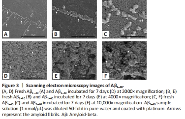
The appearances of fresh Aβ1–40 and Aβ1–40 incubated for 7 days are shown in Figure 3A–C. Prolonged incubation led to an increase in fibrillar length (Figure 3D–F).
Figure 5|Representative light photomicrograph of Congo red-stained rat retinal sections showing the effect of intravitreal administration of BDNF on Aβ1–40 deposition in the rat retina.
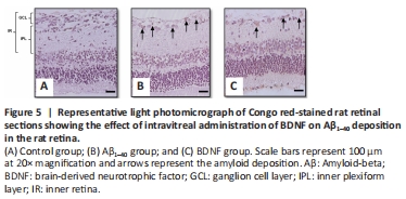
Congo red staining is part of a set of histochemical techniques used to confirm the presence of amyloid deposits based on a characteristic deep-red or salmon color. In this study, Congo red staining clearly demonstrated the presence of amyloid deposits in both the Aβ1–40- and BDNF groups (Figure 5A–C).
Figure 6|Effect of BDNF on the density of Fluoro-Gold-positive cells in Aβ1–40 exposed retina.
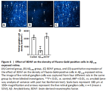
Retrograde labeling of RGCs with Fluoro-Gold showed that RGC density was 3.99-fold lower in the Aβ1–40 group than in the control group (P = 4 × 10⁻⁶). Rats that received BDNF showed 3.98-fold higher number of Fluoro-Gold-labeled RGCs than those that received Aβ1–40 (P = 4 × 10⁻⁶). The number of Fluoro-Gold-positive cells was comparable between the control and BDNF groups (Figures 6A–D).