脑损伤
-
Figure 1|Growth of primary rat BMECs in the integrin-binding array platform.
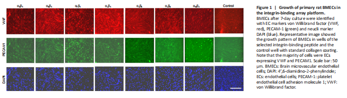
In this study, we assessed the growth of primary brain endothelial cells in our custom-designed integrin-binding array system. This array platform contained short synthetic peptides binding to 16 types of integrins, including α1β1, α2β1, α3β1, α4β1, α5β1, α6β1, α7β1, α6β4, αvβ1, αvβ3, αvβ5, αvβ6, αvβ8, αLβ2, αMβ2 and αIIbβ3, which are commonly expressed on almost all cells in vertebrates. BMECs isolated from p7 rat brain cerebral cortex were grown in the integrin-binding array plate for 7 days. Using EC cell markers including VWF and PECAM-1, as well as nuclei marker DAPI, we found about 95% of cultured cells were VWF and PECAM-1 positive, suggesting the purity of ECs with our culture method. The assessment of growth of BMECs on the integrin-binding array platform with VWF and PECAM-1 immunostaining demonstrated that most of the integrin-binding peptides supported the attachment and survival of BMECs (Figure 1).
Figure 2|Proliferation of primary rat BMECs in the integrin-binding array platform.
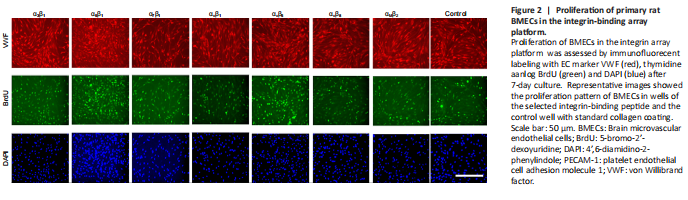
BMECs were cultured with thymidine analogue BrdU for 48 hours to assess their proliferation. Extensive VWF/BrdU double-labeled cells were found in all wells (Figure 2). Among 16 integrin-binding peptides, BMECs grown in the wells with several integrin-binding peptides including α5β1, α7β1, αvβ1, αvβ5 or αvβ8 had a significantly higher percentage of VWF/BrdU double labeled cells than cells grown in the standard collagen coated cells, among them BMECs grew in α5β1 had the highest proliferation rate (Figure 3). Subsequently, α5β1 was used in the 3D culture studies.
Figure 4|Growth of primary rat BMECs in three-dimensional culture in integrin binding peptide-conjugated hydrogels.
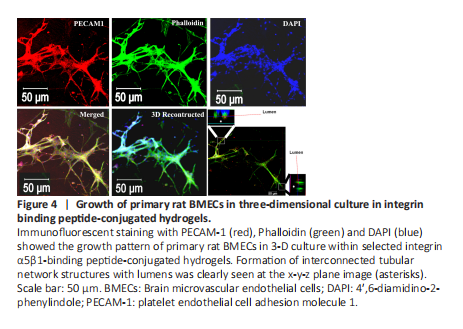
To assess the effect of integrin peptide on promotion of BMECs growth, we tested α5β1, αvβ5 and αvβ8 integrin-binding peptides for the 3D culture based on the cell proliferation study. We found α5β1 was the best one, and therefore we selected α5β1-binding peptide for the final 3D culture study. In this study, we first assessed different concentrations of α5β1 integrin binding peptide at the concentration of 0.2, 0.5, 1.0 or 1.5 μg/μL was mixed with the hydrogel and BMECs first, then the mixed components were added to the culture wells that were pre-coated with hydrogel. For the 3-D culture, the density of BMECs was at 80,000 cells/well. VEGF (0.2 μg/μL) was also added to the culture. After 7-day culture, we found that BMECs had the best growth in wells with α5β1 integrin binding peptide at 0.2 μg/μL but not in wells with higher concentrations. Taking 0.2 μg/μL as the optimal concentration for α5β1 integrin binding peptide, we then assessed different concentrations of VEGF for the growth of BMECs in 3D culture. VEGF was added to the 3D culture at 0.2, 0.5, 1.0 or 1.5 μg/μL. After 7-day culture, ECs were fixed and processed for immunostaining for PECAM-1, VWF or F-actin plus DAPI. We found that in the presence of VEGF at 1 μg/μL, BMECs had numerous sprouts-like structures from the main trunks spreading in the culture (Figure 4). The vascular-like network structures were positive for PECAM-1, F-actin and VWF, and lumen patency was assessed in x-y-z three planes with the Zen 2011 software (Figure 4). When BMECs grew in 3-D culture with α5β1-binding peptide but without VEGF or with VEGF but without integrin binding peptide, no robust tubular formation or sprouting was observed. This finding suggests that α5β1-binding peptide and VEGF work synergistically in promoting growth and interconnected and lumenized vascular-like ECs network formation of BMECs in the 3-D culture.