脑损伤
-
Figure 1|Injection and distribution of fluorescent microspheres.
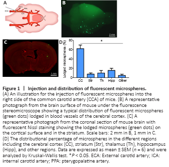
Six hours after the operation all mice were anesthetized again by intraperitoneal injection of pentobarbital sodium (50 mg/kg) and transcardially perfused with saline followed with 4% paraformaldehyde in 0.1 M phosphate buffer (PB, pH 7.4). The brain was dissected out and post-fixed in 4% paraformaldehyde in PB for 2 hours, then cryoprotected overnight in 25% sucrose. Fluorescent microspheres on the cerebral cortex were observed over the brain surface (Figure 1) using a fluorescence stereomicroscope (MVX10; Olympus, Tokyo, Japan). After examining the whole brain, each was cut into 80 μm-thick coronal sections with a freezing microtome (Thermo, Microm International GmbH, Walldorf, Germany) and the sections were collected in order in a six-hole Petri dish with 0.1 M PB (pH 7.4). The sites of lodged microspheres and microinfarcts in the brain were assessed according to a previous study (Paxinos and Franklin, 2001).
Figure 2|The vascular alteration induced by fluorescent microspheres.

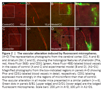
Figure 3|The neuronal degeneration in the region of microinfarcts.
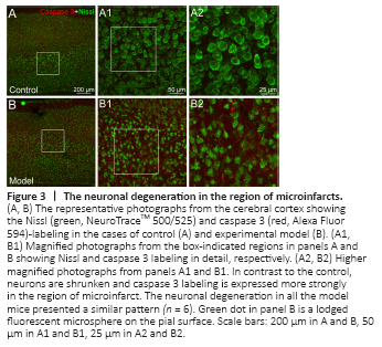
Figure 4|The astroglial and microglial activation in different regions of microinfarcts.
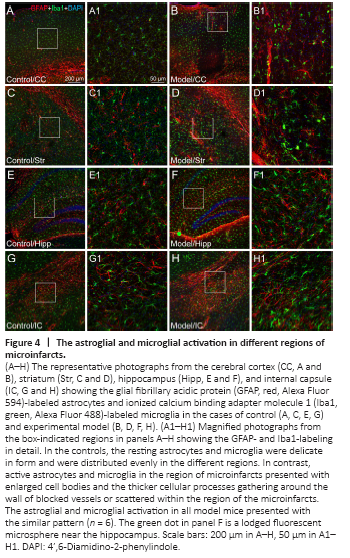
Six hours after intracarotid injection, the fluorescent microspheres were directly observed on the brain surface under the fluorescence stereomicroscope (Figure 1B). Additionally, the distribution of fluorescent microspheres was further mapped in the coronal sections of the brain (Figure 1C). Microspheres were located in the arterioles, predominately in the cortex but also in the striatum, thalamus, hippocampus and other regions in the cerebral hemisphere ipsilateral to the side of injection (Figures 1–4). Microinfarcts were detected in the tissue near where the microspheres lodged in the occluded arterioles or their downstream branches (Figures 2–4). Corresponding to the locations of microspheres in the brain, the microinfarcts were scattered in the cortex, striatum, thalamus, hippocampus and other regions. In the cortex, microinfarcts were found in typical wedge shape or column following the lodged microspheres in the penetrating arteriole at the pial surface (Figures 2–4). Microinfarcts in other cerebral regions varied in size and shape (Figures 2–4). A similar pattern of the distribution of fluorescent microspheres and microinfarcts was found in each microinfarct modelled mouse.
Figure 5|A simplified illustration of vascular and cellular alteration of microinfarct induced by a lodged fluorescent microsphere in the cerebral blood vessel.
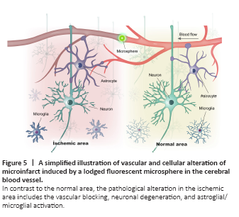
In this study, we described the pathological properties of multifocal microinfarcts in the mouse brain induced by the intracarotid injection of fluorescent microspheres. Taking advantage of this experimental model, our results provide histochemical views of the multicellular changes in the regions of cerebral microinfarcts at an early stage (Figure 5). This model of ischemic stroke may be beneficial in understanding the underlying mechanisms of multifocal microinfarcts and has the potential to assess novel therapeutic interventions of ischemic stroke.