周围神经损伤
-
Figure 1|Change in mNSS and Morris water maze performance in GCI/RI rats.
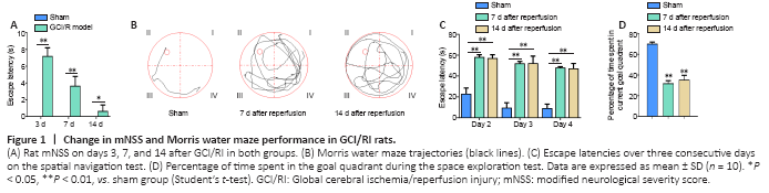
The mNSS was higher in the model group than in the sham group on days 3, 7, and 14 (3 days: t = –17.678, P < 0.01; 7 days: t = –8.000, P < 0.01; 14 days: t = –2.828, P < 0.05), and gradually decreased with time (F(2,24) =107.918; P < 0.001; Figure 1A). Analysis showed that the mNSS scores at day 3 were higher than those on days 7 or 14 (P < 0.001), and the mNSS scores on days 7 and 14 continued to decline.
The swimming paths of the rats in the sham group were smoother than those in the other three groups (Figure 1B). On the spatial navigation test, the escape latency was greater in the model group than in the sham group for three consecutive days starting from day 7 (2 days: t = ?3.137, P < 0.01; 3 days: t = ?5.509, P < 0.01; 4 days: t = ?7.628, P < 0.01) and from day 14 (2 days: t = ?6.195, P < 0.01; 3 days:, t = ?13.561, P < 0.01; 4 days: t = ?15.305, P < 0.01; Figure 1C). Moreover, time spent in the goal quadrant was less in the model group than in the sham group on days 7 and 14 (day 7: t = 37.484, P < 0.01; day 14: t = 21.934, P < 0.01; Figure 1D).
Figure 3|Apoptosis-related factors, BDNF, and TrkB expression levels in the hippocampus on days 3, 7, and 14 after GCI/RI.

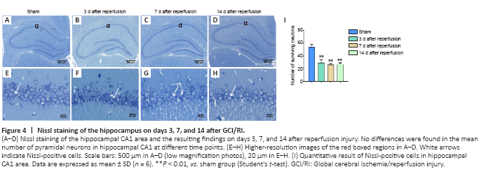
Figure 4|Nissl staining of the hippocampus on days 3, 7, and 14 after GCI/RI.
Caspase-3 expression in hippocampus on days 3 (t = ?4.342, P = 0.012), 7 (t = ?12.487, P = 0.006), and 14 (t = ?4.21, P = 0.014) after reperfusion was significantly higher than that in the sham group. Moreover, hippocampal Bcl-2/Bax levels were significantly lower in the model group than in the sham group on each day (day 3: t = 13.920, P < 0.01; day 7: t = 8.922, P < 0.05; day 14: t = 12.529, P < 0.01; Figure 3A?C).
In the sham group, CA1 neurons were large and round with uniformly colored cytoplasm. Further, the Nissl bodies around the cytoplasm were uniformly blue with normal morphology and a full shape (Figure 4A). Conversely, on days 3, 7, and 14 after injury, CA1 neurons in the model group were deformed and showed karyopyknosis, blurry cell outlines, loose arrangement, and enlarged spaces. Moreover, very few neurons showed normal morphology (Figure 4A?H). CA1 neurons were counted under high magnification. The results revealed fewer hippocampal neurons in the model group on days 3 (t = 11.215, P < 0.01), 7 (t = 10.56, P < 0.01), and 14 (t = 12.411, P < 0.01) after reperfusion than in the sham group (Figure 4I).
BDNF expression 3 days (t = ?4.195, P < 0.05) after reperfusion was significantly higher in the model group than in the sham group. BDNF expression 7 days (t = 3.944, P < 0.05) and 14 days (t = 2.697, P < 0.05) after reperfusion was significantly lower in the model group than in the sham group (Figure 3A and D). TrkB expression was lower in the model group than in the sham group on all 3 time points (day 3: t = 42.203, P < 0.01; day 7: t = 39.299, P < 0.01; day 14: t = 11.462, P < 0.05; Figure 3A and E).
Figure 6|Correlation results between hippocampal/plasma BDNF or TrkB levels with the degree of hippocampal injury after GCI/RI in rats.
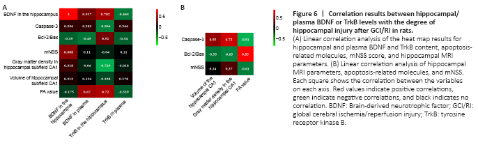
We performed linear correlation analyses between BDNF and TrkB content (hippocampal and plasma) with the mNSS, mean gray matter density of CA1, CA1 volume, FA value, hippocampal Caspase-3, Bcl-2/Bax ratio, and BDNF expression (Figure 6A). Hippocampal BDNF content correlated significantly with mNSS scores (r = 0.608; P < 0.05). Plasma BDNF did not correlate with hippocampal BDNF (r = 0.517, P = 0.104), Caspase-3 (r = 0.283, P = 0.372), CA1 mean gray matter density (r = ?0.064, P = 0.843), CA1 volume (r = 0.124, P = 0.613), mNSS (r = ?0.04, P = 0.872), or Bcl-2/Bax (r = ?0.45, P = 0.141).
Hippocampal TrkB correlated with hippocampal BDNF (r = 0.702, P < 0.05), apoptosis-related molecules (Caspase-3: r = ?0.504, P < 0.05; Bcl-2/Bax: r = 0.52, P < 0.05), mean gray matter density of CA1 (r = ?0.729, P < 0.01), and FA (r = 0.72, P < 0.01). However, plasma TrkB content did not correlate with FA value (r = ?0.329, P = 0.297), CA1 volume (r = 0.178, P = 0.579), mean gray matter density of CA1 (r = ?0.018, P = 0.957), mNSS score (r = 0.11, P = 0.743), Bcl-2/Bax (r = ?0.34, P = 0.28), Caspase-3 (r = 0.266, P = 0.403), or hippocampal BDNF (r = ?0.465, P = 0.15).
We performed linear correlation analyses between CA1 mean gray matter density, CA1 volume, and FA value, with mNSS, hippocampal Caspase-3 levels, and the Bcl-2/Bax ratio (Figure 6B). After GCI/RI, the CA1 volume and gray matter density differed from those of other subregions. Further, we found no differences in CA1 gray matter densities on days 3, 7, or 14 after reperfusion. Therefore, we only selected CA1 volume and gray matter density, as well as the hippocampal FA value, Caspase-3, Bcl-2/Bax ratio, and mNSS scores, for correlation analyses.