脊髓损伤
-
Figure 1|Serotonergic control of locomotion and pattern of serotonin fiber expression in the uninjured and injured mouse spinal cord.

The rhythmic discharge of the motorneurons that controls locomotor movements results from the activity and connections between the different spinal premotor interneurons. In this regard, neuromodulation plays a central role by modifying locomotor activity through several mechanisms: either by modulating the properties of motorneurons and CPG interneurons, or by modulating the synaptic connections between interneurons and motorneurons. It is now well established that locomotion is modulated by three monoaminergic systems that project to the spinal cord: serotonergic (5-HT), noradrenergic and dopaminergic (Pearlstein, 2013). Among those, the 5-HT system has been recognized as a powerful modulator of CPG-dependent locomotion (Ballion et al., 2002; Takakusaki et al., 2004; Ghosh and Pearse, 2014; S?awińska and Jordan, 2019). 5-HT axons observed in the spinal cord come from serotonergic neurons mostly localized in the raphe nuclei of the brainstem, including the raphe obscurus, raphe pallidus and raphe magnus. Descending brainstem 5-HT axons project then to the dorsal and ventral horn and on the intermediate area of the spinal cord (Figure 1A). Several studies in rodents have shown that locomotion is highly associated to 5-HT release in the lumbar spinal cord of rodents and the modulation of 5-HT and its receptors using agonists can produce a fictive locomotion (for review, see Ghosh and Pearse, 2014; S?awińska and Jordan, 2019). Indeed, 5-HT can regulate locomotion, rhythm and pattern of movements by modulating directly the intrinsic properties and excitability of motorneurons or indirectly through its interaction with spinal CPG interneurons to influence the amplitude and frequency of the final motor output. All these findings demonstrate that precise targeting and patterning of serotonergic projections onto thoraco-lumbar CPG networks is therefore crucial for efficient locomotion (Ghosh and Pearse, 2014; S?awińska and Jordan, 2019).
Incomplete lesions of the spinal cord can preserve part of descending axonal pathways such as 5-HT axons. The conservation of these few serotonergic fibers is of genuine importance since they have the capacity to undergo sprouting in the spinal cord with time following SCI (Saruhashi et al., 2008; Leszczyńska et al., 2015). Endogenous restoration of locomotor function observed after SCI has been shown to be partially linked to the density of 5-HT fibers sprouting of those residual serotonergic fibers (Figure 1B).
Figure 2|Sema7A is required for the proper serotonergic projections patterning in the lumbar spinal cord and for gait and locomotion functions in healthy uninjured and injured mice.
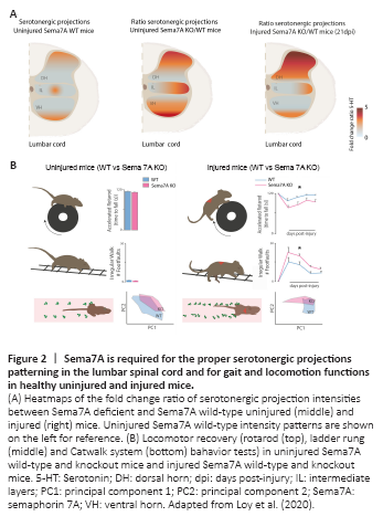
Using healthy young adult Sema7A deficient mice (2 to 3 months old), we observed that Sema7A homozygous deletion results in a significant increase of the serotonergic innervation affecting not only the dorsal and ventral horns but also the intermediate layers of the lumbar spinal cord (Loy et al., 2020; Figure2A). Despite this excess of serotonergic innervation, we did not observe an increase of contacts onto motoneurons or an increase in 5-HT2A receptors expression. In addition, we did not detect any abnormalities of locomotor and gait functions evaluated by three different behavior tests such as (i) the ladder rung test which assess skilled walking and limb placement, (ii) the Catwalk system, in which several aspects of gait and locomotion are analyzed, (iii) the rotarod test, that evaluates the balance and motor coordination. These findings support the fact that when Sema7A is constitutively deleted, there are compensatory strategies put in place to preserve locomotor function. This is not surprising as the control of locomotion also involves monoaminergic neuromodulators other than serotonin (e.g., noradrenalin and dopamine) (Pearlstein, 2013) as well as several motor tract systems, whose sprouting could to be Sema7A-independant as demonstrated in Loy et al. (2020) in the corticospinal tract. Overall, these data allowed the identification of Sema7A as a key restrictive cue for serotonergic innervation in the adult spinal cord. Then, when we challenged the spinal cord with a spinal thoracic lesion, we found that the absence of Sema7A results in an exuberant serotonergic projection to the dorsal horn of the lumbar spinal cord overtime (Figures 1B and 2A). This indicates that Sema7A also restricts the post-injury remodeling of serotonergic connectivity in the adult spinal cord. This maladaptive serotonergic patterning was paralleled by an increase of contacts onto motoneurons, impaired recovery of gait and locomotor functions as well as the appearance of signs of hindlimbs spasticity in Sema7A deficient mice (S?awińska and Jordan, 2019; Loy et al., 2020) (Figure 2B).