脑损伤
-
Figure 1|Task-state functional magnetic resonance imaging procedure.
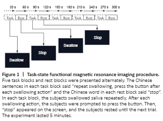
We used a block design to test brain activation related to the saliva-swallowing task. Five task blocks and five rest blocks were alternately carried out (Figure 1). Each block lasted 30 seconds. Chinese sentences and words were presented in red on a black background on a paper screen. During the scanning task, when “repeat swallowing, press the button after each swallow” appeared on the screen, the subjects swallowed saliva (lip closure, flat tongue at the bottom of the mouth, upper hyoid lift and circumpharyngeal muscle contraction). After each swallowing action, the subjects were required to press the button. Once they pressed the button, “stop” appeared on the screen, and the subjects rested for 30 seconds. During the task, the subjects kept their head motionless to concentrate on completing the swallowing task. Stimuli were presented using an image projector and a paper screen located in front of the subjects’ feet. The subjects viewed the screen through a 45° angled mirror attached to the head coil of the MRI setup. The subjects were trained before scanning to ensure their cooperation and ability to complete the task. We used a General Electric signal 3.0T magnetic resonance scanner (General Electric Company, Boston, MA, USA) with an 8-channel head coil with foam filling and earplugs to limit patient head movements and reduce noise. All subjects underwent a routine scan to identify unrelated intracranial organic lesions. The whole brain was scanned using three-dimensional T1 bravo sequences. The scanning line was consistent with the T2 fluid attenuated inversion recovery sequence. The scanning parameters were as follows: repetition time = 8.1 ms, echo time = 3.1 ms, flip angle = 90°, field of view = 30 cm × 30 cm, matrix = 300 × 300, slices = 164, thickness = 1 mm. For task state fMRI, we adopted a gradient echo planar imaging sequence. The scanning line was consistent with the T2 fluid attenuated inversion recovery sequence, and the scanning parameters were as follows: repetition time = 2000 ms, repetition time = 30 ms, flip angle = 90°, field of view = 28 cm × 28 cm, matrix = 94 × 32, slices = 40, thickness = 4 mm, space = 1 mm.
Figure 2|Functional magnetic resonance imaging showing changes in activation in individuals with Parkinson’s disease with dysphagia and healthy controls during the saliva-swallowing task.

Figure 3|Functional magnetic resonance imaging of brain activation changes in patients with Parkinson’s disease with dysphagia and healthy controls during a saliva-swallowing task.
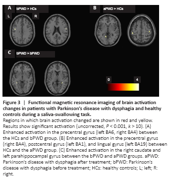
The activated brain regions in the HCs, as well as in the PWD group before versus after rTMS treatment are shown in Table 3 and Figure 2 (corrected at the cluster level of P < 0.05 with family wise error). Compared with the HCs, the PWD group had enhanced activation in the precentral gyrus (PCG; left BA6, right BA4) before rTMS treatment and enhanced activation in the PCG (right BA4), postcentral gyrus (left BA1), and lingual gyrus (left BA19) after rTMS treatment, as shown in Table 4 and Figure 3 (uncorrected, P < 0.001, k > 10).
Figure 4|Effect of repetitive transcranial magnetic stimulation on the brain regions activated during the saliva-swallowing task in individuals with Parkinson’s disease with dysphagia.
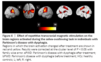
For the PWD group, activation intensity of the bilateral PCG, supplementary motor area (SMA), and cerebellum was higher after versus before rTMS, and higher than that in the HCs at both time points. The opposite was observed in the PHG, caudate, and left thalamus. Moreover, the activation intensity of the right thalamus in the PWD group was lower after rTMS versus before rTMS (Figures 4 and 5).