周围神经损伤
-
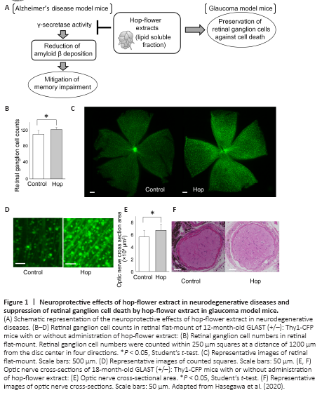
Figure 1|Neuroprotective effects of hop-flower extract in neurodegenerative diseases and suppression of retinal ganglion cell death by hop-flower extract in glaucoma model mice.
In glaucoma, a leading cause of blindness, retinal ganglion cells are progressively damaged. Intraocular pressure reduction is the only established treatment for glaucoma (Collaborative Normal-Tension Glaucoma Study Group, 1998; Vass et al., 2007). However, in some cases, visual field loss progresses despite sufficiently reduced intraocular pressure (Collaborative Normal-Tension Glaucoma Study Group, 1998; Killer and Pircher, 2018). While intraocular pressure and age are known risk factors for glaucoma (Ernest et al., 2013), the underlying mechanisms of glaucoma progression are not fully understood. Many factors that may influence glaucoma progression, including myopia and blood flow impairment, have been investigated (Marcus et al., 2011; Ernest et al., 2013). A factor that may be related to glaucoma is the concurrent occurrence of Alzheimer’s disease, which is caused by the accumulation of amyloid β (Aβ) in the brain. Glaucomatous retinal changes in patients with Alzheimer’s disease have been reported (Wang and Mao, 2021). Other studies have reported Aβ accumulation in animal models of glaucoma with ocular hypertension (Guo et al., 2007; Ito et al., 2012). Moreover, Aβ induces apoptotic retinal ganglion cell death (Guo et al., 2007). Considering that Aβ induces retinal ganglion cell death, reducing Aβ accumulation and consequently preventing retinal ganglion cell death may be a therapeutic strategy for glaucoma (Figure 1A).
To evaluate the effect of hop-flower extract over a longer period, hop-flower extract was administered orally to GLAST+/– mice starting from 1 month of age until 18 months of age (Hasegawa et al., 2020). Mice that were orally administered the hop-flower extract had ad libitum access to water containing 1 g/L of hop-flower extract, which resulted in a daily uptake of approximately 0.2 g/kg of the extract. GLAST knockout mice were cross-bred with Thy1-CFP mice, whose retinal ganglion cells express cyan fluorescent protein (Feng et al., 2000), to generate GLAST+/–:Thy1-CFP mice. Retinal ganglion cells were imaged in vivo using a scanning laser ophthalmoscope and imaged on retinal flat-mount after enucleation. The retinal ganglion cell numbers counted with the scanning laser ophthalmoscope and the retinal flat-mount were significantly higher in 12-month-old GLAST+/– mice receiving hop-flower extract than in non-treated mice (Hasegawa et al., 2020) (Figure 1B–D). The optic nerve was thicker in 18-month-old GLAST+/– mice that received hop-flower extract than in non-treated mice (Hasegawa et al., 2020) (Figure 1E and F). Immunostaining of the optic nerve of 18-month-old GLAST+/– mice did not show any difference in Aβ deposition between mice that received hop-flower extract and non-treated mice (Hasegawa et al., 2020).