神经损伤与修复
-
Figure 1|Effects of enriched environment on the body weight of offspring rats exposed to sevoflurane inhalation in the second trimester of pregnancy.
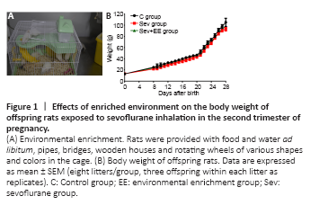
Figure 2|Flow chart of experimental procedure.
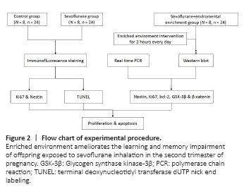
After the offspring were born, eight litters (24 offspring) from each group were randomly selected with three offspring from each litter serving as replicates. The offspring of the control and sevoflurane groups were fed normally in standard cages (BFSS19909, Nanjing Bianzhen Biotechnology Co., Ltd., Nanjing, China). The offspring of the sevoflurane + enriched environment group received 2 hours (8:00–10:00) of enriched environment intervention every day from days 8–28 after birth. For the environmental enrichment intervention, the offspring (5–6 offspring per cage) and the mother rats were put into the enriched environment cage and provided food and water ad libitum, pipes, bridges, wooden houses and rotating wheels of various shapes and colors in the cage (supplies from Grammy Pet Store, Shenyang, China; Figure 1A). We reset the objects every day to avoid repetition. After the intervention, the offspring rats and their mothers were raised in standard cages. The weight of offspring rats was measured before intervention each day. A water maze test was carried out on day 29 after birth (Figure 2).
There was no significant differences in body weight between the three groups at birth and before the beginning of the enriched environment (P > 0.05). Over time, the weight of offspring rats in the sevoflurane + environmental enrichment group increased more than that in the sevoflurane group, although the difference was not statistically significant (P > 0.05; Figure 1B).
Figure 3|Effect of enriched environment on the learning and memory impairment of offspring exposed to sevoflurane inhalation in the second trimester of pregnancy.
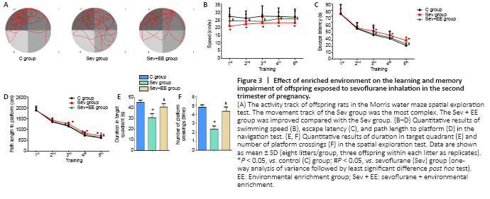
The activity track of the Morris water maze spatial exploration test for the three groups of offspring rats is shown in Figure 3A. The activity of the control group demonstrated a clear preference for the target quadrant. The sevoflurane group lacked a clear preference; the activity was evenly distributed throughout the whole region. The activity of the sevoflurane + environmental enrichment group was increased compared with that of the sevoflurane group. The swimming speed of the sevoflurane group was significantly lower than that of the control group (P < 0.05), and the swimming speed of the sevoflurane + environmental enrichment group was significantly higher than that of the sevoflurane group (P < 0.05, Figure 3B).
In the water maze directional navigation test, the escape latency of the sevoflurane group was significantly longer than that of the control group (P < 0.05), and the escape latency of the sevoflurane + environmental enrichment group was significantly shorter than that of the sevoflurane group (P < 0.05, Figure 3C). Additionally, the path length to the platform of the sevoflurane group was significantly longer compared with that of the control group (P < 0.05), and the path length to the platform of the sevoflurane + environmental enrichment group was significantly shorter compared with that of the sevoflurane group (P < 0.05, Figure 3D). In the water maze spatial exploration test, the duration in the target quadrant of the sevoflurane group was significantly shorter than that of the control group (P < 0.05), and duration in the target quadrant of the sevoflurane + environmental enrichment group was significantly increased compared with that of the sevoflurane group (P < 0.05, Figure 3E). The number of platform crossings in the sevoflurane group was significantly decreased compared with that in the control group (P < 0.05), and the number of platform crossings in the sevoflurane + environmental enrichment group was significantly increased compared with that in the sevoflurane group (P < 0.05, Figure 3F). These results suggest that an enriched environment for offspring reduces learning and memory functional impairment induced by sevoflurane inhalation by mothers in the second trimester of pregnancy.
Figure 4|Effect of enriched environment on the proliferation of hippocampal neural stem cells of offspring exposed to sevoflurane inhalation in the second trimester of pregnancy.
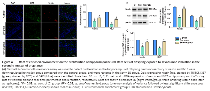
The immunoreactivity of Ki67 and nestin in the sevoflurane group were significantly lower than those in the control group, and the immunoreactivity of Ki67 and nestin in the sevoflurane + environmental enrichment group were significantly higher than those in the sevoflurane group (all P < 0.05, Figure 4A). Western blot and real-time polymerase chain reaction were used to detect the protein and mRNA expression, respectively, of nestin and Ki67 in the hippocampus of offspring rats. The protein and mRNA expression of nestin and Ki67 in the sevoflurane group were significantly lower than those in the control group (all P < 0.05), and the protein and mRNA expression of nestin and Ki67 in the sevoflurane + environmental enrichment group were significantly higher than those in the sevoflurane group (all P < 0.05; Figure 4B and C). These results suggest that an enriched environment reduces the inhibition of hippocampal neural stem cell proliferation induced by sevoflurane anesthesia.
Figure 5|Effect of enriched environment on hippocampal neural stem cells through GSK-3β pathway.
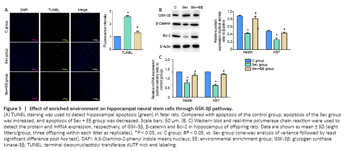
The number of TUNEL-positive cells in the sevoflurane group was significantly higher than that in the control group, and the apoptotic level in the sevoflurane + environmental enrichment group was lower than that in the sevoflurane group (P < 0.05; Figure 5A). Previous studies have shown that sevoflurane can regulate the proliferation and apoptosis of hippocampal neural stem cells by affecting the GSK-3β pathway (Wang et al., 2018a, b). The results of western blot and real-time polymerase chain reaction showed that in the hippocampus of the sevoflurane group, the protein and mRNA expression of β-catenin and Bcl-2 were significantly lower than those in the control group (P < 0.05), and the protein and mRNA expression of GSK-3β were significantly higher than those in the control group (P < 0.05). Compared with the sevoflurane group, the sevoflurane + environmental enrichment group had significantly increased protein and mRNA expression of β-catenin and Bcl-2 (P < 0.05), and significantly decreased protein and mRNA expression of GSK-3β (P < 0.05; Figure 5B and C). All of these results suggest that an enriched environment for offspring ameliorates the alterations in proliferation and apoptosis of hippocampal neural stem cells induced by sevoflurane inhalation by mothers in the second trimester of pregnancy through the GSK-3β pathway.