神经损伤与修复
-
Figure 1|Effect of SKP-SCs on cell viability and autophagy in SH-SY5Y cells under 6-OHDA intervention.

Our previous study demonstrated that SKP-SC-CM showed a cytoprotective effect in SH-SY5Y cells as measured by CCK-8 assay (Chen et al., 2020). Here, immunofluorescence showed that 6-OHDA significantly increased the intensity of LC3B immunoreactivity, where the highest intensity of LC3B was observed when cells were treated with 50 μM 6-OHDA for 12 hours (Figure 1A). This same trend was observed for the intensity of p62 (Figure 1B).
To examine the effects of SKP-SC-CM on 6-OHDA-injured cells, we measured the expression levels of TH and α-syn by western blot assay. TH is the rate-limiting enzyme in catecholamine biosynthesis, and thus is crucial in regulating the biosynthesis of dopamine (Lindenbach et al., 2017). Additionally, abnormal accumulation of α-syn protein has been reported during the progression of PD (Thomsen et al., 2021). In the present study, pretreatment with SKP-SC-CM markedly reduced α-syn expression (P < 0.05, vs. 6-OHDA group) and significantly increased TH expression (P < 0.01, vs. 6-OHDA group, Figure 1C).
We also found that compared with the control group, the 6-OHDA induction group had an increased level of autophagy, and SKP-SC-CM inhibited the increased autophagy. The LC3B-II/I ratio and the expression of p62 significantly decreased in the SKP-SC-CM group (P < 0.05, vs. 6-OHDA group, Figure 1D), which indicates that the mechanism underlying SKP-SC-CM-mediated neuroprotective effects against 6-OHDA may be associated with autophagy regulation (Figure 1E–H).
Figure 2|An autophagy agonist diminishes the neuroprotective effects by reversing autophagy inhibition by SKP-SC-CM.
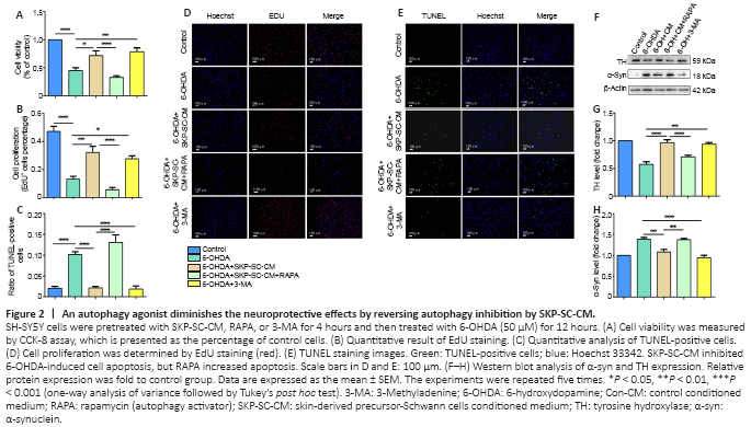
To further confirm the effects of autophagy regulation by SKP-SC-CM, the autophagy activator RAPA or autophagy inhibitor 3-MA was added 2 hours before 6-OHDA in the 6-OHDA-treated cells. As shown in Figure 2A, SKP-SC-CM increased cell viability, which was decreased after treatment with 6-OHDA (P < 0.05, vs. 6-OHDA group). The cell viability of the SKP-SC-CM + RAPA group was lower than that of the SKP-SC-CM group (P < 0.001). Pretreatment with 3-MA increased cell viability in the 6-OHDA + 3-MA group compared with that of the 6-OHDA group (P < 0.01).
SKP-SC-CM significantly increased proliferation and inhibited the 6-OHDA-induced apoptosis in SH-SY5Y cells (Figure 2B and C). Pretreatment with RAPA reduced the increased rate of proliferation (P < 0.001, vs. SKP-SC-CM group), whereas pretreatment with 3-MA increased proliferation after exposure to 6-OHDA (P < 0.05, vs. 6-OHDA group, Figure 2D). We also examined apoptosis by TUNEL assay. As shown in Figure 2E, apoptosis in the SKP-SC-CM treatment group was decreased compared with that in the 6-OHDA group (P < 0.001), which was similar to the changes seen in the 6-OHDA + 3-MA group (P < 0.001, vs. 6-OHDA group). In contrast, apoptosis in the SKP-SC-CM + RAPA group was significantly increased compared with apoptosis after SKP-SC-CM treatment alone (P < 0.001).
We also measured the expression levels of TH and α-syn. The TH level increased after pretreatment with SKP-SC-CM or the autophagy inhibitor 3-MA compared with the 6-OHDA group (P < 0.01 for both comparisons, Figure 2F and G). Similarly, SKP-SC-CM or 3-MA decreased the expression of α-syn in comparison with 6-OHDA treatment alone. RAPA reversed the decrease in α-syn expression caused by pretreatment with SKP-SC-CM (P < 0.01, vs. SKP-SC-CM group, Figure 2F and H).Figure 3|RAPA weakens the protective effects of SKP-SC-CM by activating autophagy in 6-OHDA-induced SH-SY5Y cells.
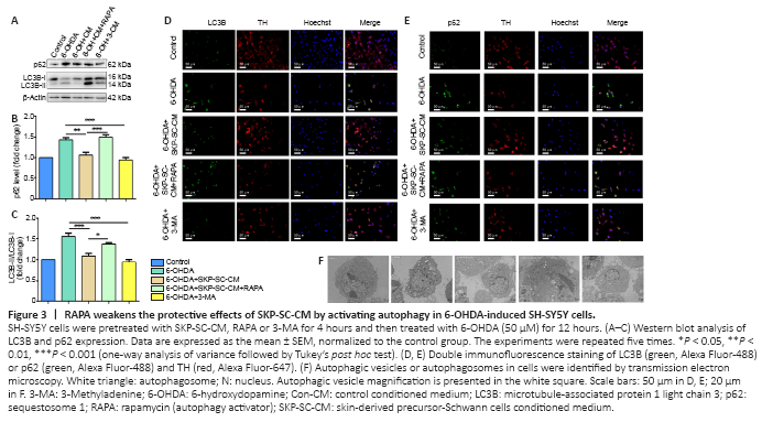
To investigate the relationship between autophagocytosis and the neuroprotective effect of SKP-SC-CM, we examined autophagy levels. The LC3B-II/I ratio and expression of p62 significantly decreased after treatment with SKP-SC-CM (P < 0.01, vs. 6-OHDA group) or 3-MA (P < 0.001, vs. 6-OHDA group). Moreover, treatment with RAPA increased autophagy (P < 0.05, vs. SKP-SC-CM group, Figure 3A–C). Immunofluorescence staining of LC3B and p62 were both upregulated in the 6-OHDA group compared with the control group. The addition of SKP-SC-CM or 3-MA to the 6-OHDA-treated cells decreased the intensity of both LC3B and p62 (Figure 3D and E).
Autophagosomes, the hallmark structures of autophagy, form characteristic double-membrane vesicles (Kwon et al., 2019). We found an increasing number of autophagosomes after the treatment of 6-OHDA. This result suggests that 6-OHDA injury may be related to the overactivation of autophagy. In addition, transmission electron microscopy analysis demonstrated that SKP-SC-CM or 3-MA in 6-OHDA-injured SH-SY5Y cells decreased the number of autophagosomes compared with 6-OHDA treatment alone (Figure 3F). These results suggested that SKP-SC-CM downregulated excessive autophagy in the 6-OHDA-induced cell model.Figure 4|SKP-SC-CM regulates 6-OHDA-induced autophagy by upregulating the PI3K/AKT/mTOR pathway in SH-SY5Y cells.
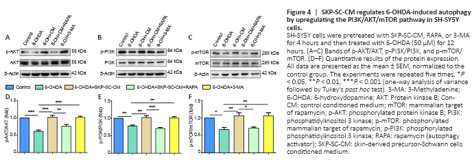
The PI3K/AKT pathway exhibits prosurvival and antiapoptotic effects in neurodegenerative disease (Rai et al., 2019). The mTOR complex 1 (mTORC1) is a major negative regulator of autophagy, and the PI3K/AKT pathway is a main upstream modulator of mTORC1 (Heras-Sandoval et al., 2014). In the present study, p-PI3K and p-AKT expression was increased after SKP-SC-CM treatment compared with those in the 6-OHDA group (P < 0.001 and P < 0.01, respectively). Additionally, these increases were diminished by treatment with RAPA (P < 0.01 and P < 0.001, respectively, vs. SKP-SC-CM group). Notably, the 3-MA group similarly increased p-PI3K and p-AKT expression (P < 0.001 and P < 0.01, respectively, vs. 6-OHDA group, Figure 4A and B).
Finally, we examined the levels of phosphorylated mTOR (p-mTOR) and total mTOR protein using western blot assay. As Figure 4C shows, the ratio of p-mTOR/mTOR in the SKP-SC-CM and 3-MA pretreatment groups was increased compared with that in the 6-OHDA group (P < 0.01 for both comparisons). After the addition of RAPA to SH-SY5Y cells, the p-mTOR/mTOR level decreased (P < 0.05, vs. SKP-SC-CM group). These results suggested that SKP-SC-CM protected against 6-OHDA-induced neurotoxicity by regulating autophagy via the PI3K/AKT/mTOR pathway (Figure 4D–F).