脑损伤
-
Figure 1|Rectal temperature during the global cerebral ischemia model procedure.
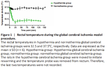
This study used male animals only because female animals have higher estrogen levels that protect them from infection. Eighteen male Sprague-Dawley rats, specific-pathogen-free level, 9–11 weeks old, weighing 320–350 g, were randomized into three groups. The hypothermia GI group (n = 6) was maintained at 31–33°C to establish the GI model. The temperature probe was inserted into the rectum and the rats were placed on a cooling/warming blanket (CMA 150, Carnegie Medicine, Stockholm, Sweden) that was connected to a thermostatically controlled system (CMA 150, Carnegie Medicine) that maintained the temperature using feedback regulation. During a 25-minute cooling period, alcohol was sprayed on the fur of rats to reduce body temperature. The hypothermia period was maintained for 150 minutes, after which the rats were warmed for 30 minutes, and then maintained at normal temperature until scanned. The normothermia GI group (n = 6) was maintained at 37–37.5°C using cooling/warming blankets with the same system to establish the GI model. Duration of normothermia was maintained for 150 minutes (Figure 1). The sham group (n = 6) was maintained at 37–37.5°C and subjected to a median incision in the neck but without induction of GI model.
Figure 2|The hypometabolic regions in the global cerebral ischemia model rats by PET.

PET results showed the hypometabolic regions of whole brain were significantly smaller in the hypothermic GI group when compared with those in the normo-thermic GI group (Figure 2A and B).
Figure 3|The hypometabolic regions in anterior forebrain and Ts of the mesocircuit in the global cerebral ischemia model rats.

When compared with the normothermic GI group, the hypothermia GI group also had significantly smaller hypometabolic regions in the Ts and prefrontal-cortex (PFC) and primary sensory areas (Figure 3A–D). These results indicate that hypothermia selectively protects the anterior forebrain (cortical) and Ts (subcortical part) of the mesocircuit under GI.
Figure 4|Difference of brain injury on MRI T2 images between normothermia GI (A) and hypothermia GI (B) groups.

The results from the MRI T2 scans show obvious injury to the Ts region in the normothermia GI group (Figure 4A). There was almost minimal brain injury in Ts region in the hypothermia GI group (Figure 4B).
点击此处查看全文