脑损伤
-
Figure 2| Identification of exosomes derived from BMSCs overexpressing miR-23b.

MiR-23b was upregulated in the BMSC group transfected with lentivirus carrying miR-23b (LV-miR-23b) (Figure 2A). The transmission electron microscope revealed that the exosomes derived from the BMSCs exhibited a round cup-shaped and complete structure (Figure 2B). Western blot analysis further identified positive expression of the exosomal biomarkers CD63, TSG101, and CD81 in the exosomes derived from the BMSCs (Figure 2C). As shown in Figure 2D, we observed a higher miR-23b level in exosomes from BMSCs transfected with miR-23b (exomiR-23b), suggesting that the overexpressed miR-23b could be delivered to BMSC-derived exosomes.
Figure 3| BMSC-exosomal miR-23b improves behavioral functions and reduces ICH-induced brain injury.

To explore the effects of the BMSC-derived exosomes carrying miR-23b in ICH rats, the same volume of the exomiR-23b, exomiR-NC, or PBS was injected, and an increased miR-23b level was found in the perihematomal brain tissue in the exomiR-23b group (Figure 3A). To observe the distribution of the BMSC exosomes, we labeled the exosomes with PKH67 green fluorescence and the microglia/macrophages with Iba1 staining. We found that the exosomes were delivered successfully into the microglia/macrophages in the perihematomal region (Figure 3B). The neurological deficits were evaluated using the corner tests and limb placement assessments. ICH induction increased the frequency of right turns on days 1, 3, and 7 compared with that in the sham group, and the increase was mitigated by exomiR-23b administration on day 7 (Figure 3C). Similar neurological function improvements were observed using limb placement tests (Figure 3D). The brain water content of the ipsilateral hemisphere showed a significant increase after ICH induction, which was attenuated by exomiR-NC or exomiR-23b administration (Figure 3E). Brain tissue morphology in ICH rats was evaluated using H&E staining. Non-regularly arranged neuronal cells, deep staining, and a decreased number of nuclei with neutrophil infiltration were observed in the ICH group compared with those in the sham group. However, the large pathological deteriorations induced by ICH were attenuated in both exomiR-NC and exomiR-23b groups. The fewest histological impairments were observed in rats that received exomiR-23b administration (Figure 3F).
Figure 4| BMSC-exosomal miR-23b inhibits the amount of microglia/macrophages and neuronal apoptosis in brain tissues.
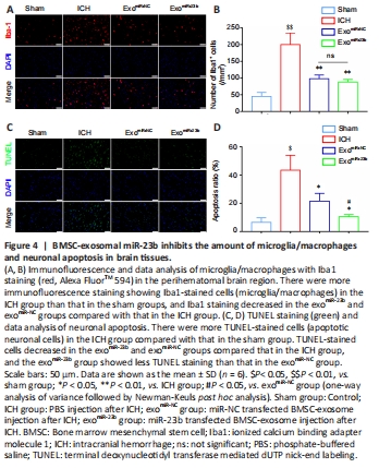
Iba1 fluorescence staining for microglia/macrophages was performed to observe the inflammation in ICH rats. The amount of Iba1-positive cells was elevated after ICH induction but decreased by exomiR-NC and exomiR-23b injection, whereas there was no significant difference between exomiR-NC and exomiR-23b injections (Figure 4A and B). Cell apoptosis was assessed via the amount of TUNEL-positive cells. As depicted in Figure 4C and D, administration of exomiR-23b or exomiR-NC reduced cell death induced by ICH, and the exomiR-23b group further alleviated cell apoptosis compared with that in the exomiR-NC group.
Figure 5| BMSC-exosomal miR-23b inhibits oxidative stress in brain tissues.

ROS staining was used to estimate the oxidative stress level. The ROS level in the perihematomal brain tissue was increased after ICH induction compared with that in the sham group, but decreased after exomiR-23b administration compared with that in the ICH group (Figure 5A and B). We evaluated lipid peroxidation by estimating the MDA level, and we found that the MDA level was increased after ICH induction compared with that in the sham group, but it was decreased by exomiR-23b compared with that in the ICH group (Figure 5C). Major antioxidant enzyme SOD activity in brain tissue was elevated in the exomiR-23b group compared with that in the exo miR-NC or ICH group (Figure 5D). GSH and GSSG maintain a dynamic balance, and a reduced GSH/GSSG ratio means that there is elevated oxidative stress. The ICH group exhibited a decreased GSH/GSSG ratio compared with that in the sham group. However, exomiR-23b administration significantly increased the ratio compared with that in the ICH group (Figure 5E). Collectively, the results confirmed the antioxidant effect of exosomal miR-23b.
Figure 7| Exosomal miR-23b inhibits oxidative stress in microglia BV2 cells in vitro.

We stimulated microglia BV2 cells with 60 μM hemin for 24 hours to mimic ICH conditions in vitro, and exosomes were incubated separately with hemin-stimulated microglia BV2 cells; PBS incubation was set as the control. The results showed that the miR-23b level in the exomiR-23b group was higher than that in the other groups (Figure 7A). Oxidative stress was evaluated using ROS, MDA, SOD, and GSH/GSSG levels. BV2 cells treated with 60 μM hemin induced the increase in intracellular ROS, which was reversed by the treatment with the antioxidant NAC, confirming that the increase in ROS was induced by hemin (Figure 7B and C). Additionally, exomiR-23b could significantly decrease the ROS levels induced by hemin (Figure 7B and C). Similar results were also observed in MDA measurements (Figure 7D). SOD levels were markedly decreased by hemin stimulation, and exomiR-23b improved the SOD levels compared with that in the hemin group (Figure 7E). Furthermore, the decrease in the GSH/GSSG ratio induced by hemin was reversed by exomiR-23b administration (Figure 7F).