周围神经损伤
-
Figure 4|Number of axons and myelin sheath thickness in the tibial nerve in rats.
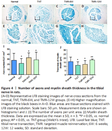
Compared with the normal group, axon numbers were significantly decreased in the TNT group (P < 0.05; Figure 4A–I). In comparison with the TNT group, axon numbers were significantly increased in the TMR-6W and TMR-12W groups (P < 0.05). There was no statistically significant difference in axon numbers between the TMR-6W and TMR-12W groups (P > 0.05; Figure 4A–I).
Compared with the normal group, the myelin sheath was significantly thinner in the TNT group (P < 0.05; Figure 4A–H, J) and in the TMR-6W and TMR-12W groups (P < 0.05; Figure 4A–H, J). However, 12 weeks after TMR, the myelin sheath was significantly thicker in the TMR-12W group than in the TNT group (P < 0.05).
Figure 5|Expression of S100-B (a marker for myelin sheaths of nerves) on the operated side of rats.

S100-B immunoreactivity on the operated side of rats was significantly increased in the TNT group compared with the normal group (P < 0.05) and was significantly decreased in the TMR-6W and TMR-12W groups compared with the TNT group (P < 0.05; Figure 5).
Figure 6|Nissl staining of cross sections from the lumbosacral enlargements of the spinal cords of rats.
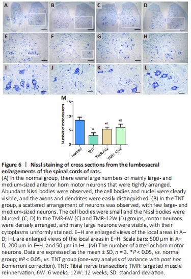
The number of motor neurons was significantly lower in the TNT group than in the normal group (P < 0.05; Figure 6). The number of motor neurons was significantly higher in the TMR-6W group than in the TNT group (P < 0.05). In comparison with the TMR-6W group, the number of motor neurons in the TMR-12W group was increased, but there was no statistically significant difference between the two groups (P > 0.05; Figure 6).
Figure 7|Choline acetyltransferase immunoreactivity in the spinal cords of rats.

The ChAT-positive cells were arranged in a disordered manner, and the number of cholinergic neurons were significantly reduced in the TNT group compared with the normal group (P < 0.05, Figure 7I). ChAT immunoreactivity was also significantly decreased in the TNT group compared with the normal group (P < 0.05). In comparison with the TNT group, the number of cholinergic neurons in the spinal cord was significantly increased (P < 0.05) and ChAT immunoreactivity was significantly enhanced (P < 0.05; Figure 7J) in the TMR-6W and TMR-12W groups.
Figure 8|Synaptophysin expression in motor neurons in the anterior horn of the spinal cord in rats.
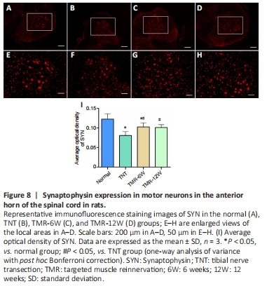
Immunofluorescence staining revealed strong SYN immunoreactivity in motor neurons in the anterior horn of the spinal cord of rats in the normal group. Compared with the normal group, SYN-positive cells were arranged in a disordered manner and SYN immunoreactivity was significantly reduced in the TNT group (P < 0.05; Figure 8). Compared with the TNT group, SYN immunoreactivity in motor neurons in the anterior horn of the spinal cord was significantly enhanced in the TMR-6W and TMR-12W groups (P < 0.05; Figure 8).