脑损伤
-
Figure 3|Characterization of TPM4-targeted Ag/Au hollow nanoshells@SiO2 nanoparticles.
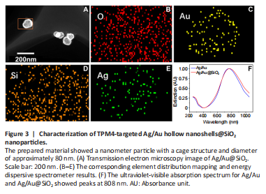
The prepared material showed a nanometer particle with a cage structure and diameter of approximately 80 nm (Figure 3). Based on a distribution mapping diagram of the elements O, Au, Si, and Ag, the elements were evenly distributed throughout the nanoparticles. The ultraviolet-visible absorption spectra for Ag/Au and Ag/Au@SiO2 showed peaks at 808 nm.
Figure 6|Pathological analysis of cerebral cortex supplied by the left middle cerebral artery (affected side).
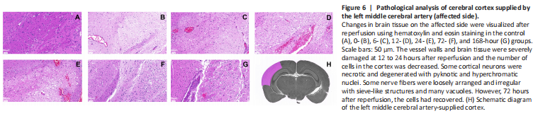
Figure 7|Tropomyosin 4 (TPM4) immunohistochemical analysis of cortex supplied by the left middle cerebral artery (affected side).
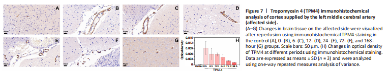
After photoacoustic examination at each time point, the mice were sacrificed and their brain tissue extracted for histological examination. Brain tissue damage was assessed on hematoxylin and eosin-stained slides (Figure 6). TPM4 immunohistochemical staining showed tissue damage on the affected side was severe in the 1-, 24-, and 168-hour groups, mild in the 12- and 72-hour groups, and moderate in the 6-hour group (Figure 7).