脑损伤
-
Figure 2|Time course and location of Piezo1 expression following ICH.
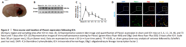
Piezo1 protein levels were detected by western blotting to explore the changes in Piezo1 expression following ICH in experiment I (Figure 2A). Compared with the sham group, Piezo1 expression was significantly higher 3 to 48 hours after ICH (P < 0.05) and peaked at 3 hours (Figure 2B and C). After the 48-hour time point, the Piezo1 expression declined rapidly, and within 72 hours of the cerebral hemorrhage, the symptoms of the mice in the ICH group were similar to those seen in the sham group (P > 0.05; Figure 2B and C). Immunofluorescence staining performed 3 hours after ICH demonstrated that Piezo1-positive cells also expressed Olig2, an oligodendrocyte marker (Joseph et al., 2016; Figure 2D).
Figure 4|The effects of Dooku1 on white matter injury following ICH.
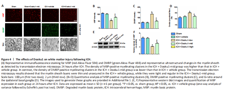
Approximately 30% of central nervous system myelin is made up of MBP, which is used as a protein marker of demyelination following ICH (Joseph et al., 2016). In experiment III, immunofluorescence staining showed myelinating clusters red or green complete circles. Fewer MBP-positive myelinating clusters were observed after ICH injury than in the sham mice (P < 0.05, vs. sham group; Figure 4A), and treatment with Dooku1 (10 mg/kg) significantly increased the number of MBP-positive myelinating clusters at 24 hours after ICH (P < 0.05, vs. ICH + vehicle group, Figure 4A and B). dMBP is produced by myelin degradation (Xia et al., 2019) and is used as a biomarker of demyelination. At 24 hours after ICH, the number of dMBP-positive myelinating clusters was significantly increased (P < 0.05, vs. sham group; Figure 4A and C), and treatment with Dooku1 (10 mg/kg) markedly decreased the number of dMBP-positive myelinating clusters (P < 0.05, vs. ICH + vehicle group; Figure 4A and C). Western blot analysis indicated that MBP expression levels were also significantly lower at 24 hours after ICH (P < 0.05, vs. sham group; Figure 4E and F). Treatment with Dooku1 (10 mg/kg) markedly increased MBP expression (P < 0.05, vs. ICH + vehicle group; Figure 4E and F).
Next, transmission electron microscopy was used to observe the ultrastructure of myelinated nerve fibers in the ipsilateral basal ganglia following ICH. The myelin sheath layers were tight and regular in the sham group. At 24 hours after ICH, the myelin sheath layers were thin and unsecured, leading to a significant increase in the G-ratio (P < 0.05, vs. sham group; Figure 4A and D). However, treatment with Dooku1 (10 mg/kg) alleviated the degree of demyelination and significantly decreased the G-ratio (P < 0.05, vs. ICH + vehicle group; Figure 4A and D).
Figure 5|The effects of Dooku1 on oligodendrocyte apoptosis following ICH.
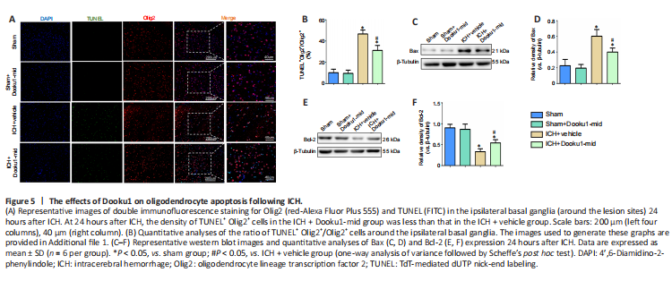
In experiment III, the role of Piezo1 in oligodendrocyte apoptosis in the ipsilateral basal ganglia region was assessed by administering Dooku1 (10 mg/kg) and assessing cell apoptosis by TUNEL assay 24 hours after ICH. At 24 hours after ICH, the number of TUNEL-positive oligodendrocytes was significantly greater in the ICH groups than in the sham group (P < 0.05, vs. sham group; Figure 5A and B), while the number of TUNEL-positive oligodendrocytes in the ICH + Dooku1-mid group was lower than that in the ICH + vehicle group (P < 0.05, vs. ICH + vehicle group; Figure 5A and B).
Bax and Bcl-2 are both key factors in cell apoptosis (Edlich, 2018). Bax expression was significantly increased and Bcl-2 expression was significantly decreased around the hematoma in the ipsilateral basal ganglia after ICH (both P < 0.05, vs. sham group; Figure 5C–F), while treatment with Dooku1 (10 mg/kg) significantly upregulated Bax expression and downregulated Bcl-2 expression (both P < 0.05, vs. ICH + vehicle group; Figure 5C–F).
Figure 6|The effects of Dooku1 on GRP78 expression in oligodendrocytes following ICH.
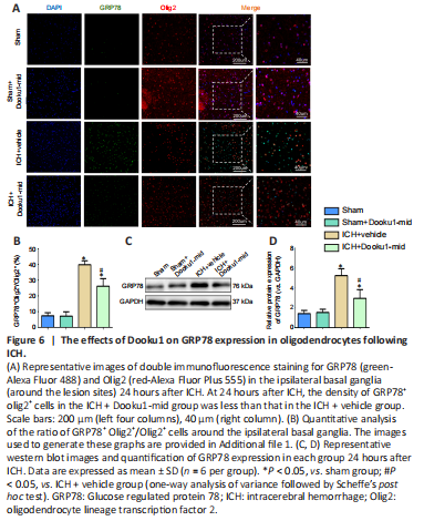
GRP78 is an important indicator of ER stress (Sano and Reed, 2013). In experiment III, at 24 hours after ICH, the number of GRP78-positive, olig2-positive cells was significantly higher in the ICH groups than in the sham group (P < 0.05). Compared with the ICH + vehicle group, the number of GRP78-positive oligodendrocytes in the ICH + Dooku1-mid group was significantly lower (P < 0.05; Figure 6A and B). The western blot results also showed significant elevation of GRP78 expression 24 hours after ICH compared with the sham group (P < 0.05). Treatment with Dooku1 (10 mg/kg) significantly decreased GRP78 expression (P < 0.05, vs. ICH + vehicle group; Figure 6C and D).
Figure 7|The effects of Dooku1 on CHOP expression in oligodendrocytes following ICH.
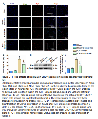
CHOP is also important for ER stress-induced apoptosis (Sano and Reed, 2013) and is known to be involved in ICH-induced apoptosis (Liew et al., 2019). At 24 hours after ICH, the number of CHOP-positive, olig2-positive cells was significantly higher in the ICH groups compared with the sham group (P < 0.05). Compared with the ICH + vehicle group, the number of GRP78-positive oligodendrocytes in the ICH + Dooku1-mid group was significantly lower (P < 0.05; Figures 7A and B). The western blot results also showed a significant increase in CHOP expression at 24 hours after ICH (P < 0.05) compared with the sham group. Treatment with Dooku1 (10 mg/kg) significantly decreased CHOP expression (P < 0.05, vs. ICH + vehicle group; Figure 7C and D).