脊髓损伤
-
Figure 1|Effects of PBM on mitochondrial dynamics within neurons in spinal cord at 1, 7, and 14 days after SCI.
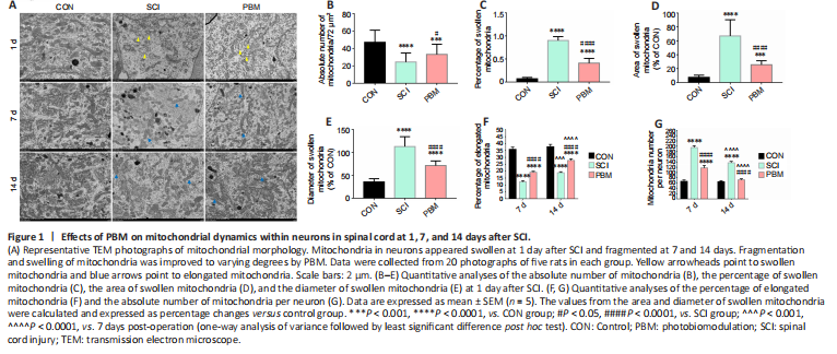
To explore the effects of PBM on the dynamic tubulation of mitochondria, TEM was used to examine mitochondrial morphology in individual neurons from rats at 1, 7 and 14 days postsurgery (Figure 1). Representative TEM images showed that mitochondria were present in dynamic networks in control rats (Figure 1A). At 1 day, mitochondrial alterations in the neurons from SCI rats were characterized by decreased mitochondrial number, increased mitochondrial area and cristae loss, and the percentage of swollen mitochondria in SCI rats was higher than that in the control animals. Treatment with PBM restored the alterations in mitochondrial number, mitochondrial volume, cristae loss and the percentage of swollen mitochondria to some extent, though the measures did not reach the levels of the control group (Figure 1A–E and Additional Figure 2). At 7 and 14 days, the absolute number of mitochondria per neuron in SCI rats was significantly higher than that in control rats, and the majority of mitochondria exhibited short and round appearance (Figure 1A, F and G). PBM treatment attenuated the decrease in the percentage of elongated mitochondria (Figure 1F) and the increase in mitochondria number (Figure 1G) at 7 and 14 days. Interestingly, SCI rats at 14 days post-surgery had a higher percentage of elongated mitochondria (Figure 1F) and a lower number of mitochondria (Figure 1G) than those at 7 days, and PBM treatment for 14 consecutive days had a larger effect than treatment for 7 consecutive days (Figure 1F and G). Data of the PBM treatment groups did not reach the control group levels except for mitochondria number at 14 days post-surgery (Figure 1F and G).
Figure 4|Effects of PBM on neuronal injury at 1, 7 and 14 days after SCI.
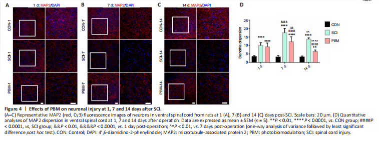
To assess the impact of PBM on neuronal injury, MAP2 dispersion was examined to assess the degree of neuronal injury. Confocal fluorescence images indicated that the neurons in the gray matter of the normal ventral spinal cord were tightly arranged with low MAP2 dispersion (Figure 4A–C). SCI significantly increased MAP2 dispersion at 1 (Figure 4A), 7 (Figure 4B) and 14 (Figure 4C) days after injury. SCI rats at 7 days had a higher level of MAP2 dispersion than those at 1 day (Figure 4D). PBM did not improve MAP2 dispersion at 1 day (Figure 4A), and PBM treatment for 7 and 14 consecutive days decreased MAP2 dispersion, but MAP2 dispersion did not reach the levels of the controls (Figure 4D). TUNEL staining confirmed that PBM improved apoptosis at 14 days after SCI (Additional Figure 3). Rats with PBM treatment at 7 days had a higher level of MAP2 dispersion compared with rats with PBM treatment at 1 day (Figure 4D). In addition, MAP2 dispersion was lower in the SCI rats at 14 days after operation than that at 7 days, suggesting self-repair of neuronal injury (Figure 4D). The neurons of ventral spinal cord around the injured area from rats with 14-day PBM had substantially lower MAP2 dispersion than those with 7-day PBM (Figure 4D).