视神经损伤
-
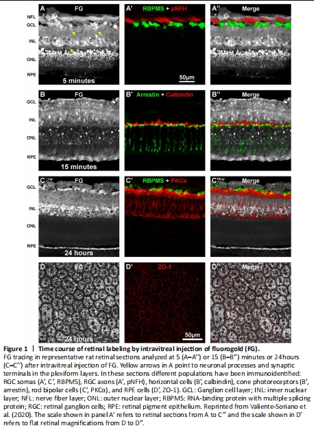
Figure 1|Time course of retinal labeling by intravitreal injection of fluorogold (FG).
In this perspective, we revise the course of FG tracing in intact retinas (Figure 1).
Intravitreal tracing with FG is a fairly quick process and 5 minutes after its administration, the somas of neurons in the ganglion cell layer (GCL), inner nuclear layer (INL) and outer nuclear layer (ONL) are already traced (Figure 1A–A’’). In the plexiform layers, neuronal processes and synaptic terminals are also delineated (Figure 1A, yellow arrows). In the GCL, some traced-somas are RGCs since they co-localize with the RNA-binding protein with multiple splicing protein (RBPMS) (Rodriguez et al., 2014), while the rest must be displaced amacrine cells which, together with RGCs are the neurons located in the GCL. This is the best time point to visualize RGC axons. Indeed, RGC axons are now beautifully traced (Figure 1A–A’’) but lose the tracing rather quickly. In fact, RGC axons have lost most of the tracing 15 minutes after FG administration (Figure 1B–B’’) and are no longer traced after 6 hours (Figure 1 in Valiente-Soriano et al., 2020). The same course is observed for the processes in the plexiform layers.
Fifteen minutes of tracing labels more somas in the ONL where photoreceptors (rods and cones) lay, reaching their outer segments and finally, the RPE (Figure 1B–B’’). The tracing in the ONL and the outer segments is also transient, diminishing gradually between 1 and 24 hours (Figure 1 in Valiente-Soriano et al., 2020) when no outer segments and only few and faintly traced photoreceptor somas are observed (Figure 1C–C”).
In sections analyzed 24 hours after the tracing, the RPE is clearly labeled but, as said above, the outer retina is mainly devoid of FG. Interestingly, many somas in the inner retina remain labeled (see RGCs in the GCL and rod-bipolar cells in the INL immunodetected in Figure 1C).
The disappearance of the tracer from the photoreceptors is due to one of the most important functions of the RPE (Sparrow et al., 2010), the phagocytosis of shed photoreceptors’ outer segment membrane. This phagocytosis explains why the tracer disappears from the outer retina but remains in the inner regions. Because the tracing of RPE occurs when the RPE cells phagocytose the outer segments of photoreceptors, this approach is very useful to study the functionality of the RPE in models of retinal degeneration (Valiente-Soriano et al., 2020).
The accumulation of FG in the RPE cells is better observed in flat mounts than in sagittal sections (compare panels C and D in Figure 1). Immunodetection of zonula occludens, a marker of tight junction protein 1 that delimits the morphology of the RPE cells (Zech et al., 1998; Georgiadis et al., 2010), shows that FG accumulates inside the cytoplasm avoiding the nuclei and extracellular spaces (Figure 1D–D’’). Thus FG tracing reveals the characteristic honey-comb morphology of the RPE cells and their one or two nuclei per cell (Bonilha, 2008; Bhatia et al., 2016).