周围神经损伤
-
Figure 1|Proliferative activity and NGF and IL-1β secretion in Schwann cells and macrophages under different media.

According to the EdU results at 24 and 48 hours, groups C and D had stronger Schwann cell proliferation ability than groups A and B, and group D had the strongest proliferation ability. These results suggest that 2.0 ng/mL IL-1β promoted Schwann cell proliferation (Figure 1A). In the proliferation activity test of macrophages, the strongest macrophage proliferative activity was in group H. Conditioned medium of Schwann cells activated by 2.0 ng/mL IL-1β promoted macrophage proliferation (Figure 1B). ELISA was used to detect NGF levels in the different groups, and showed that of the four groups, group D had the highest NGF level in Schwann cells (Figure 1C). IL-1β levels in groups F and H were significantly higher than those in groups E and G. Conditioned medium of macrophages activated by nerve homogenate and conditioned medium of Schwann cells activated by 2.0 ng/mL IL-1β both led to high IL-1β expression in macrophages (Figure 1D).
Figure 4|The bioactive artificial nerve promotes Schwann cell proliferation in the injured area after sciatic nerve injury.】
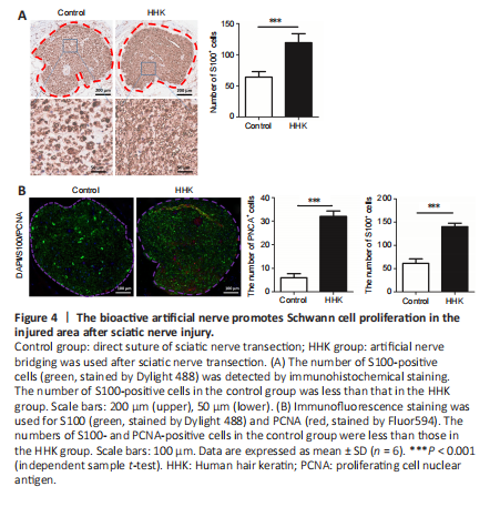
Immunohistochemical staining showed that the number of S100-positive cells was significantly greater in the HHK group than in the control group (Figure 4A). Immunofluorescence also showed that the number of PCNA-positive cells was higher in the HHK group than in the control group (Figure 4B). The results indicated that the artificial nerve significantly promoted Schwann cell proliferation.
Figure 5|The bioactive artificial nerve promotes NGF secretion in sciatic nerve under in situ hybridization.

In situ hybridization was used to detect the expression of NGF, an important factor in nerve repair. NGF expression in the HHK group was much higher than that in the control group, suggesting that the artificial nerve promoted NGF secretion and thus accelerated nerve repair (Figure 5).
Figure 6|The bioactive artificial nerve affects nerve repair factors in injured sciatic nerve.
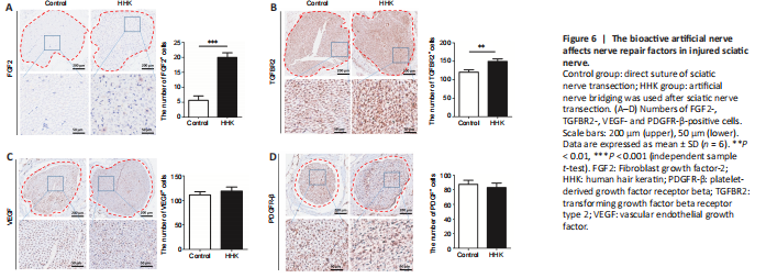
Several important proteins related to nerve repair were detected by immunohistochemical staining. The numbers of FGF2- and TGFBR2-positive cells were significantly higher in the HHK group than those in the control group (Figure 6). The artificial nerve had no significant effect on the numbers of VEGF- and PDGFR-β-positive cells. The results showed that the mechanism of promoting neural repair may be related to activation of the TGF signaling pathway and upregulation of FGF2 secretion.