脑损伤
-
Figure 4|The bFGF-chitosan gel activated neural stem/progenitor cells.
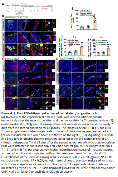
Seven days after the stroke cavity was filled with bFGF-chitosan gel, a significantly greater number of nestin-positive cells was observed in the stroke cavity compared with the stroke only sand lesion control (LC) group (P < 0.05; Figure 4A, B, and E).
No DCX+ cells were observed in the stroke cavity in the stroke only (14 days after stroke) or LC (7 days after aspiration). In comparison, 7 days after bFGF-chitosan gel application, more DCX+/BrdU+ cells were observed in the SVZ, and some even migrated to the area around the stroke cavity (P < 0.05; Figure 4C and F). At 14 days after aspiration, the neurogenesis provoked by stroke had almost ceased in the stroke only and LC groups, whereas in the bFGF-chitosan gel group, many DCX+/BrdU+ cells were observed in the SVZ, and more such cells had migrated towards the stroke cavity (P < 0.05; Figure 4D and F).
Figure 5|Generation and maturation of neurons in the stroke cavity after application of the bFGF-chitosan gel.
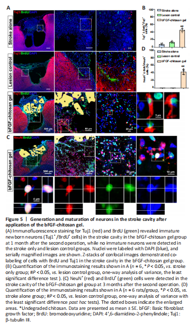
After ischemic stroke, bFGF-chitosan gel strongly activated DCX+/BrdU+ cells migrate to the damaged area. However, whether these activated cells could differentiate into neurons remained unclear. At 28 days after bFGF-chitosan gel application, many Tuj1+/BrdU+ immature neurons were observed in and around the stroke cavity, while almost no Tuj1+/BrdU+ cells were observed in the stroke cavity in the stroke only and LC groups (P < 0.05; Figure 5A and B). Three months after bFGF-chitosan gel application, NeuN+/BrdU+ cells were observed in the stroke cavity, but none were observed in the stroke cavity of the stroke only and LC groups (Figure 5C and D). These results suggest that bFGF-chitosan gel facilitates neuronal generation in the stroke cavity in the short term and promotes the survival and maturation of these neurons in the long term.
Figure 6|The bFGF-chitosan gel facilitates angiogenesis and blood vessel maturation in and around the stroke cavity.

Angiogenesis is a critical event during neural regeneration after ischemic stroke. After stroke, reconstruction of the local micro-circulating environment may prolong the survival of relevant tissues including neurons and ultimately improve the efficacy of rehabilitation of stroke patients (Slevin et al., 2006). Nih et al. (2018) reported that neuronal survival after ischemic stroke was correlated with increased blood supply. To investigate whether bFGF-chitosan gel facilitates angiogenesis after photothrombotic occlusion, we used Glut-1 to label vascular endothelial cells. In the stroke only group, at 7–14 days after stroke, the stroke cavity was empty and devoid of blood supply, with almost no blood vessels remaining. In comparison, at 7 days after bFGF-chitosan gel application in the bFGF-chitosan gel group, many newborn vascular endothelial cells (Glut-1+/BrdU+ cells) were observed in the stroke cavity, together with an greater percentage of vascular area compared with the stroke only and LC groups (P < 0.05, Figure 6C). At 14 days after bFGF-chitosan gel application, we observed similar results (Figure 6A and D). These findings suggest that bFGF-chitosan gel facilitates the long-term regeneration of vascular endothelial cells in the stroke cavity.
To investigate whether these vascular endothelial cells could develop into functional vascular networks, we assessed vascular maturity 1 and 3 months after bFGF-chitosan gel application. During angiogenesis, pericyte coverage is considered an indicator of vascular maturity and function (Zhang et al., 2012). Thus, we used platelet-derived growth factor receptor beta (Pdgfr-β) to label pericytes to assess vascular maturity (Zhang et al., 2012). Three months after bFGF-chitosan gel application, Glut-1/BrdU/Pdgfr-β–triple positive mature, functional vessels were observed in the stroke cavity (Figure 6B), suggesting that bFGF-chitosan gel could not only facilitates vascular endothelial cell generation in the short term, but also promotes the maturation and function of these vessels in the long term.