脑损伤
-
Figure 6|Effects of an miR-9-5p inhibitor and L-cysteine on brain tissue loss at 3 days after HI.
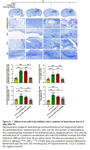
Nissl bodies are basophilic clumps and particles in neurons. Nissl bodies can be used as a marker of the functional state of neurons because its shape, number and distribution vary. The Nissl staining results indicated that L-cysteine increased the amount of Nissl bodies in the injured prefrontal cortex, hippocampal CA1, CA3, and dentate gyrus. This effect was significantly reversed by miR-9-5p inhibitor in the prefrontal cortex, hippocampal CA1, and CA3 regions (Figure 6). These results indicate that L-cysteine significantly reversed the reduction of nissl bodies after HI injury, but this effect was partially inhibited after treatment with L-cysteine combined with miR-9-5p inhibitor.
Figure 7|Effects of an miR-9-5p inhibitor and L-cysteine on cell apoptosis at 3 days after HI.
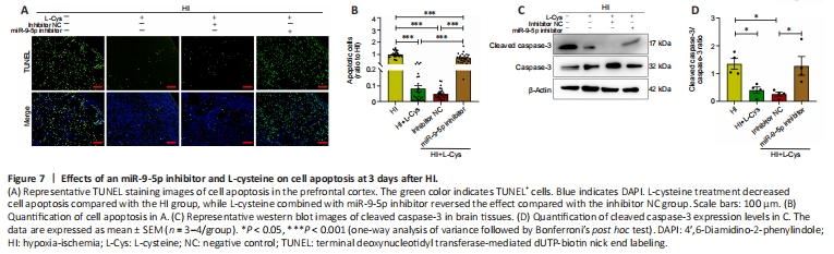
Quantitative analysis of the TUNEL staining results showed that, compared with the HI group, the injured hemisphere exhibited fewer apoptotic cells after L-cysteine administration at 3 days after HI injury. Similarly, miR-9-5p inhibitor partially reversed the protective effect of L-cysteine (Figure 7A and B). In addition, L-cysteine treatment reduced the cleaved caspase-3/caspase-3 ratio on the injured side compared with the HI group at 3 days after HI insult. This protective effect of L-cysteine on the cleaved caspase-3/caspase-3 ratio was blocked by miR-9-5p inhibitor (Figure 7C and D). These results indicate that the antiapoptotic effect of L-cysteine after HI injury was partially inhibited after miR-9-5p inhibitor administration.
Figure 8|Effects of an miR-9-5p inhibitor and L-cysteine on microglial activation in the prefrontal cortex at 3 days after HI.
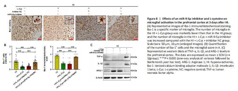
As expected, the L-Cys + miR-9-5p inhibitor group exhibited a significantly increased number of Iba-1+ cells compared with L-Cys + miR-9-5p inhibitor NC group. Furthermore, Iba-1+ cells on the injured side in the HI group were amoeboid-like, and their appearance and morphology improved substantially after L-cysteine administration and were close to those seen in resting state at 3 days after HI insult. L-Cys + miR-9-5p inhibitor partially blocked the protective effect of L-cysteine on microglial activation (Figure 8A and B).
The western blot assay results showed that L-cysteine treatment visibly decreased the expression of tumor necrosis factor alpha (TNF-α) and interleukin 1beta (IL-1β), while increasing the expression of arginase-1 (ARG-1). The combination of L-cysteine and miR-9-5p inhibitor up-regulated TNF-α and IL-1β expression, while down-regulating ARG-1 expression, compared with L-Cys + inhibitor NC treatment group at 3 days after HI injury (Figure 8C and Additional Figure 6). These findings indicate that the anti-inflammatory effect of L-cysteine after HI injury was partially inhibited by miR-9-5p inhibitor administration.