脑损伤
-
Figure 3| AAV-shEndorepellin-GFP brain transduction increases perfused small vessel density in CCI mice.
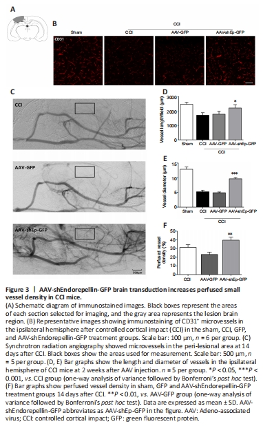
Figure 4| AAV-shEndorepellin-GFP brain transduction promotes angiogenesis 14 days after CCI.

CD31+ endothelial cells have been used as indicators of angiogenesis via microvessel immunostaining (Zhou et al., 2019). In this study, we used CD31+ endothelial cells to determine whether downregulation of endorepellin promotes angiogenesis in the peri-lesional area. Two weeks after CCI, immunostaining showed that the number and diameter of CD31+ cells in the peri-lesional area were greater in AAV-shEndorepellin-GFP mice compared with CCI mice (P < 0.05; Figure 3A, B and D). A significant increase in the diameter of CD31+ cells was also observed in AAV-shEndorepellin-GFP mice (P < 0.001; Figure 3E). Additionally, downregulation of endorepellin led to a greater number of CD31+/Ki-67+ cells in the peri-lesional area, compared with CCI mice (P < 0.01; Figure 4A and B). These results are consistent with an inhibitory effect of endorepellin on endothelial cell proliferation. To identify the factors responsible for the increased amount of CD31+ microvessels, pro-angiogenic growth factor expression within the peri-lesional brain area was examined by western blotting at two weeks after CCI. The expression levels of both VEGF and Ang-1 were significantly greater in AAV-shEndorepellin-GFP mice compared with CCI mice (P < 0.05; Figure 4B–E).
The results so far indicate that downregulation of endorepellin promotes angiogenesis after CCI. To determine if the newly formed microvessels are functional, the perfused vascular density in the peri-lesional area was quantified from SRA images obtained 14 days after injury. Perfused vessel density was higher in AAV-shEndorepellin-GFP mice compared with AAV-GFP mice (P < 0.01; Figure 3C and F). This indicates that the newly formed microvessels induced by endorepellin downregulation are functional.
Figure 5|Endorepellin downregulation improves tube formation of human umbilical vein endothelial cells.
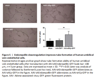
The effect of endorepellin on the ability of HUVECs to form tubes was investigated after transduction with AAV-shEndorepellin-GFP. The results showed that endorepellin inhibition promoted tube formation (Figure 5).