视神经损伤
-
Figure 1|Histopathological changes in the retina of normal control, complete Freund’s adjuvant (CFA)-only, and experimental autoimmune uveoretinitis (EAU) rats.

Under microscopic observation, the presence of round cells was confirmed in the ciliary body (Figure 1C) and retina (Figure 1F) of EAU rats, whereas these cells were not observed in normal controls (Figure 1A and D) or rats administered CFA only (Figure 1B and E). Numerous round cells (inflammatory cells) were detected in the anterior and posterior chambers near the ciliary body (Figure 1C). Furthermore, retinal detachment was confirmed in some EAU rats (data not shown).
Figure 2|Western blot analysis of osteopontin (OPN) in the retina of normal control, CFA-only, and EAU rats.

Western blot analysis confirmed the upregulation of OPN in the EAU rats compared with normal control and CFA-only rats (Figure 2). The levels of both the OPN precursor (predicted size 66 kDa) and OPN cleavage product (predicted size 25–55 kDa) were increased in the ocular tissue of EAU rats compared with normal control and CFA-only rats (Figure 2A), with significant increases in the retina (both P < 0.05) (Figure 2B).
Figure 3|Immunohistochemical staining of OPN and CD44 in the ciliary body of normal control, CFA-only, and EAU rats.
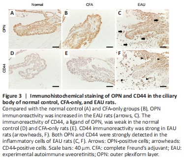
Next, we determined the OPN expression pattern in the eye. OPN immunoreactivity was detected in the ciliary body in the normal control and CFA-only groups (Figure 3A and B) and was markedly increased in the round cells around the ciliary body in EAU rats (Figure 3C). CD44, which interacts with OPN, was also upregulated in the ciliary body of EAU rats (Figure 3F) compared with the normal control and CFA-only groups (Figure 3D and E).
Figure 4|Immunohistochemical staining of OPN in the retina of the normal control, CFA-only, and EAU rats.

Furthermore, OPN was localized in the RGC layer, inner plexiform layer, inner nucleus layer, and outer plexiform layer in the normal control and CFA-only groups (Figure 4A and B). The intensity of OPN immunoreactivity was increased in all layers of the retina in EAU rats (Figure 4C).
Figure 5|Double immunofluorescence staining of OPN and ionized calcium binding adaptor molecule 1 (Iba1) or glutamine synthetase (GS) in the ciliary body and retina of normal control and EAU rats.
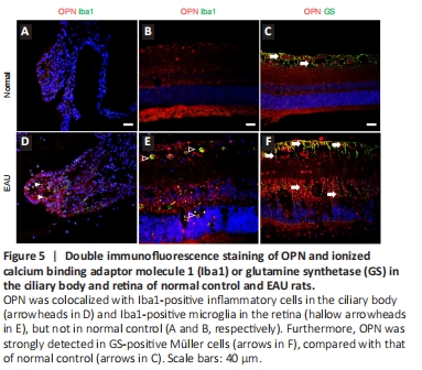
To investigate the phenotype of OPN-expressing cells in the ciliary body and retina, we performed double fluorescence staining in the eyes of normal control and EAU rats. OPN immunoreactivity was colocalized with Iba1-positive inflammatory cells in the ciliary body of EAU rats (Figure 5D), but not in normal control rats (Figure 5A). Furthermore, OPN was detected strongly in Iba1-positive microglia (Figure 5E) and glutamine synthetase-positive Müller cells in the retina (Figure 5F), compared with those of normal control rats (Figure 5B and C, respectively).