脑损伤
-
Figure 4|Tat-CX3CL1 (357–395aa) inhibits microglia polarization to an M1 phenotype 24 hours after middle cerebral artery occlusion.

PCR analysis of cytokine production showed that Tat-CX3CL1 (357–395aa) inhibited the production of M1-type pro-inflammatory factors (iNOS, TNF-α, and IL-1β) (Figure 3A–C) and increased the levels of M2-type anti-inflammatory factors (IL-10, CD163, and VEGF) 24 hours after stroke (Figure 3D–F). Immunofluorescence assay of M1-type microglia (Iba1 and CD68 double-staining) in the cortex and striatum around the infarct area after 24 hours of reperfusion showed that the number of M1 microglia was significantly decreased in the Tat-CX3CL1 (357–395aa) group, not only in cortex but also in the striatum (Figure 4A–D). These data suggest that Tat-CX3CL1 (357–395aa) affects the microglia M1/M2 phenotype switch during ischemic stroke.
Figure 7| Tat-CX3CL1 (357–395aa) reduced white matter injury after MCAO.
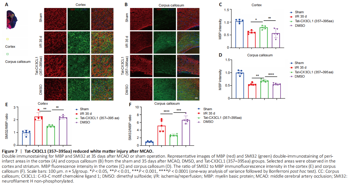
White matter injury after stroke can lead to cognitive deficit, neuroinflammation, demyelination, and axon degeneration (Rosenzweig and Carmichael, 2015; Shi et al., 2015; Wang et al., 2016). To explore whether Tat-CX3CL1 (357–395aa) could mitigate cognitive dysfunction, we performed a Morris water maze test and found that the peptide reduced escape latency and increased the time spent in the target quadrant, but it did not affect swimming speed (Figure 6A–D). Next, we evaluated whether Tat-CX3CL1 (357–395aa) could improve white matter integrity in stroke by double-staining for MBP and neurofilament H non-phosphorylated (SMI32) to evaluate white matter lesions. The immunofluorescence intensity of MBP staining was decreased in the corpus callosum and cortex at 35 days after MCAO (Figure 7A–D). However, Tat-CX3CL1 (357–395aa) markedly inhibited myelin loss. The SMI32/MBP ratio was used to assess damage to the myelin sheath and white matter. The results showed that the SMI32/MBP ratio in corpus callosum and cortex regions increased markedly in the I/R and DMSO groups compared with the sham group, but decreased in the Tat-CX3CL1 (357–395aa) group compared with the I/R and DMSO groups (Figure 7A, B, E, and F).