脊髓损伤
-
Figure 2|Motor and histopathologic changes in the mouse model of SCI.
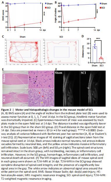
To confirm the successful establishment of the mouse model of SCI using an SCI impactor, motor function was assessed by behavioral tests (BMS score, inclined plate test, and open field test) at 1, 3, 7, and 14 dpi (Figure 2A–D). Hindlimb paralysis immediately developed in the SCI group, but did not in the sham group. Mouse spinal cords were collected for sagittal sectioning at 14 dpi, and HE staining (Figure 2E) was used to observe histopathologic changes. In the sham group, the spinal cord structures remained intact without hemorrhage, necrosis, or inflammatory cell infiltration. In contrast, the SCI group was characterized by rupture of spinal cord structures, accompanied by hemorrhage, inflammatory cell infiltration, and neuronal loss. T2-weighted MR images of sagittal slices of mouse spinal cord in each group are shown at 14 dpi in Figure 2F. A disruption of the continuity of the spinal cord structures can be seen in the MR images. There is a significant hypointense area at the lesion, indicating the presence of stale hemorrhage, and scattered hyperintense areas surrounding the lesion, indicating edema formation. Therefore, a persistent inflammatory response existed after SCI.