神经退行性病
-
Figure 1|Emodin alleviates the clinical signs and degree of inflammation and demyelination of experimental autoimmune encephalomyelitis rats.
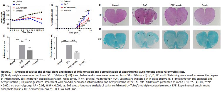
Decreased body weight and increased neurobehavioral scores indicate an increase in EAE severity. We recorded body weights and neurobehavioral scores from D0 to D14 (Figure 1). The body weights of all groups decreased from D0 to D3 and then began to increase (Figure 1A). From D10, the body weights of the control and emodin groups continued to increase, whereas the body weights of the EAE and EAE + emodin groups began to decrease. The body weights of the EAE + emodin group were between those of the control and EAE groups. There was a significant difference in body weights between the EAE + emodin and EAE groups from D4 onwards, suggesting that emodin can alleviate body weight loss in EAE rats. Both the EAE + emodin and EAE groups exhibited onset of disease symptoms on D9 and reached the peak of disease at D14, indicating that emodin did not delay the onset of EAE (Figure 1B). From D10, the neurobehavioral scores of the EAE + emodin group were decreased compared with those of the EAE group, implying that emodin alleviated neurobehavioral deficits in EAE rats.
EAE rats were sacrificed on D14 (the peak of the disease) for HE and LFB staining to assess histopathological changes in the spinal cord (Figure 1C and D). HE staining of the lumbar enlargement of the spinal cord revealed clear inflammatory cell infiltration in EAE rats, while the degree of infiltration in the EAE + emodin group was markedly reduced (Figure 1C and E). LFB staining of the lumbar enlargement of the spinal cord showed that the degree of demyelination in the EAE + emodin group was markedly reduced compared with that in the EAE group (Figure 1D and F). Collectively, these results demonstrate that emodin alleviated inflammatory cell infiltration and demyelination in an EAE rat model.
Figure 3|Emodin inhibits microglial aggregation and activation in the lumbar enlargements of experimental autoimmune encephalomyelitis rats and attenuates inflammation in BV2 cells.
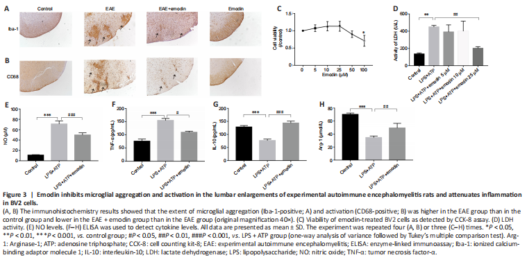
Iba-1 and CD68 levels in the lumbar enlargement of the spinal cord were assessed by immunohistochemistry. Iba-1 is mainly expressed in macrophages and reflects the degree of microglial aggregation. CD68 plays a role in the phagocytic activity of tissue macrophages and indicates the microglial M1 phenotype and degree of microglial activation (Brown and Neher, 2010). We observed a clear increase in Iba-1 and CD68 levels in the EAE group compared with those in the control group, whereas treatment with emodin inhibited this increase (Figure 3A and B).
We next performed a series of in vitro experiments to further investigate the effect of emodin on microglial activation. The results of the cell counting kit-8 assay showed a concentration-dependent effect of emodin on BV2 cell viability: cell viability increased with treatment with up to 50 μM emodin and decreased at concentrations greater than 50 μM (Figure 3C). LDH analysis demonstrated that emodin (25 μM) markedly attenuated LDH activity in cell culture supernatants from BV2 cells induced with LPS + ATP (Figure 3D). In subsequent experiments, 25 μM emodin was used as the standard treatment concentration. Follow-up ELISA and NO assays showed a marked decrease in the activation of pro-inflammatory factors (tumor necrosis factor (TNF)-α, and NO) in BV2 cells pretreated with emodin (Figure 3E and F) and a clear increase in the expression of anti-inflammatory cytokines (interleukin (IL)-10, and arginase-1) (Figure 3G and H). These findings suggest that emodin can inhibit microglial activation.