神经损伤与修复
-
Figure 1|High-dose biotin neither improves peripheral nerve regeneration nor elevates malonyl-coenzyme A (CoA) levels.
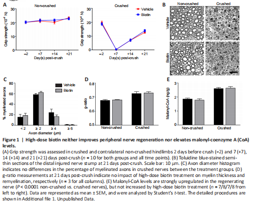
To this end, we performed sciatic nerve crush in wild-type C57BL/6J mice treated with 60 mg/kg biotin (corresponding to human equivalent dose of 300 mg per day in MS patients (Sedel et al., 2016) via daily intraperitoneal injection, starting immediately after crush injury was introduced. Animals were subjected to clinical testing by grip strength analysis of the left (non-crushed) and right (crushed) hindlimbs at 2 days before and 7, 14 and 21 days post-crush (Figure 1A). We did not observe any improvement of grip strength under HDB treatment. Coherently, histological analyses performed at 21 days post-crush (Figure 1B) revealed that HDB did not affect the distribution of myelinated axons (Figure 1C) and, most importantly, did not improve myelin thickness as determined by g-ratio measurements (Figure 1D). At 14 days post-crush, during the phase of ongoing remyelination, we prepared sciatic nerve homogenates to quantify the ACC enzymatic product malonyl-CoA, a building block for fatty acid synthesis. In line with elevated metabolic activity in the regenerating nerve, we were able to detect highly significant increases in malonyl-CoA levels in crushed nerves as compared to non-crushed controls (Figure 1E). However, there was no additional improvement under HDB treatment, indicating that the proposed mechanism of HDB driving ACC activity and thus malonyl-CoA formation is not present to a measurable extent. Of note, HDB treatment was nevertheless well tolerated by the animals and did not affect weight (Figure 2A) or basic hematological parameters (Figure 2B–G).