脑损伤
-
Figure 2|Expression of death-associated protein kinase-1 and tau in the hippocampus.
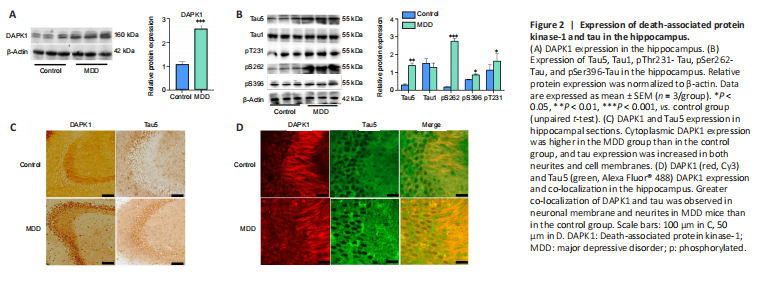
To assess the causes of the decline in cognitive function observed in the MDD mice, we analyzed the expression of proteins in various regions of the hippocampus by western blotting, immunohistochemistry, and immunofluorescence (Figure 2A–D). DAPK1 expression in the hippocampus was higher in the MDD group than that in the control group, and the protein was abnormally distributed (both P < 0.001, vs. control group; Figure 2A and C). Normally, DAPK1 is found in the neurites of neurons, and it is only expressed at very low levels in the cytoplasm and membrane (Figure 2C). However, in MDD mice, DAPK1 distribution was altered. In addition to being expressed in neurites, its expression in the cytoplasm was significantly increased (Figure 2C). Tau hyperphosphorylation is closely associated with cognitive dysfunction in AD (Duan et al., 2013; Kim et al., 2014). The western blot results showed that total tau expression (P < 0.01) and tau phosphorylation at Ser262 (P < 0.05), Thr231 (P < 0.01), and Ser396 (P < 0.001) in the hippocampal CA3 area were higher in the MDD group than in the control group, while non-phosphorylated tau (Tau1) expression levels were similar between these two groups (P > 0.05; Figure 2B). Immunohistochemistry analysis showed that DAPK1 and tau levels in the hippocampal CA3 area of MDD mice were increased in both neurites and cell membranes (Figure 2C). DAPK1 co-localization with Tau5 was increased in the hippocampal CA3 area, as demonstrated by immunofluorescence staining, indicating that these two proteins are functionally correlated (Figure 2D). These results suggest that the cognitive dysfunction observed in MDD mice may be related to increased DAPK1 expression and tau hyperphosphorylation.
Figure 6|Effect of citalopram on the expression of death-associated protein kinase-1, Tau5, pSer262-Tau, and pSer396-Tau in the hippocampi of MDD mice.

To assess whether the mechanism by which antidepressants improve cognitive function is related to DAPK1, immunoblotting was used to detect DAPK1 expression in the hippocampus. In MDD mice, DAPK1 expression was lower in the hippocampus after treatment with the antidepressant (P < 0.05; Figure 6A and B). In addition, total tau expression (P < 0.01, vs. MDD + normal saline group) and tau phosphorylation at Ser262 (P < 0.001, vs. MDD + normal saline group) and Ser396 (P < 0.05, vs. MDD + normal saline group) were lower after antidepressant treatment (Figure 6A and B), indicating that the reduction in DAPK1 expression and tau phosphorylation may be involved in the improvement in cognitive dysfunction seen in MDD mice treated with citalopram. Immunohistochemical staining showed that Ser396-phosphorylated tau was concentrated on the cell membranes in MDD mice, and that the neurites of hippocampal neurons were shorter in MDD mice than in control mice (Figure 6C). After treatment with antidepressants, pS396-Tau expression in the cell membrane was decreased (Figure 6C). Immunofluorescence experiments further showed that co-localization of neuronal membrane and neurites in the hippocampal CA3 area was increased in MDD mice, whereas their co-localization was significantly reduced in drug-treated mice (Figure 6D). Thus, tau expression may be altered by DAPK1, which may play a role in cognitive dysfunction in MDD.