周围神经损伤
-
Figure 2|Time-course microscopic and quantitative results of peripheral nerve regeneration through the conduit.
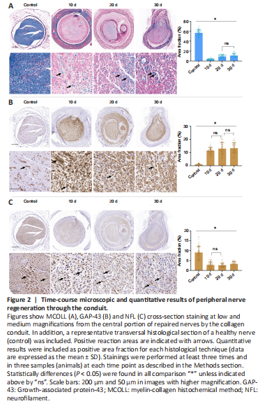
The time-course histological analyses showed an active regeneration process of the nerve gaps repaired with the collagen conduit from 10 days onwards (Figure 2). The regeneration process was observed in the conduit at all time points studied, and small newly-formed fascicles were detected. Furthermore, no signs of inflammatory or adverse reaction were observed in any animal evaluated.
Figure 3|Time-course microscopic results of the lumbar section of the spinal cord after peripheral nerve damage and repair.
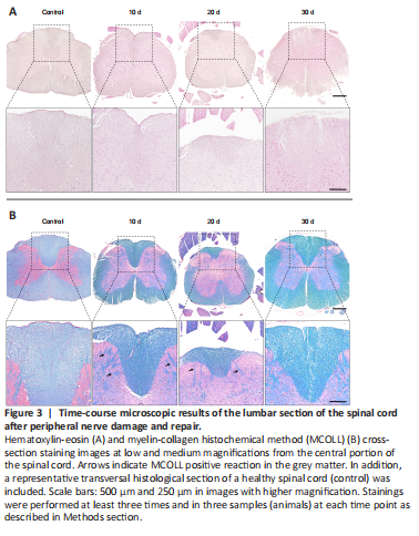
Spinal cord changes during nerve regeneration were evaluated by hematoxylin-eosin staining and MCOLL histochemical method (Figure 3). In this study, the transversal section at low magnification revealed the complete cytoarchitecture of the spinal cord, and the white and grey substances were remarkably different as shown by MCOLL method. Hematoxylin-eosin staining results revealed no apparent changes at the cellular (neuronal and glial) level or fiber organization. However, the MCOLL technique at higher magnification suggested slight changes in the structural organization of the posterior cord, as compared with the control group (Figure 3).
Figure 4|Immunohistochemical and quantitative evaluation of GAP-43 and NFL expression in the spinal cord after peripheral nerve damage and repair.
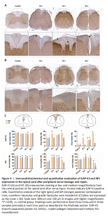
Interestingly, important differences were observed in GAP-43 expression at the spinal cord level during sciatic nerve regeneration process (Figure 4A and C). Histology revealed a considerable increase in GAP-43 immunoreaction in the grey and white matters, as compared with the control group, from 10 days onward. Surprisingly, these changes were especially evident in the white matter of the posterior columns. At 10 days post-repair, GAP-43 was found to be located in certain regions of the left cuneiform fasciculus, differing from the pattern found in the control group, where it was just observed in the descending pathway of the corticospinal tract of the posterior funiculus. Furthermore, GAP-43 staining at 20 days post-repair was consistent in most spinal cord areas, with a similar expression pattern in the cuneiform fasciculus and in the corticospinal tract (Figure 4A and C). At 30 days post-repair, GAP-43 expression exhibited an overall homogenous distribution, with a remarkable increase in the left posterior cord, covering the cuneiform fasciculus and the gracile fasciculus. These results were corroborated by the quantitative area fraction analyses conducted in the white posterior columns which revealed remarkable statistical differences in operated animals as compared with the control group (P < 0.05). Interestingly, the increase in GAP-43 area fraction positive reaction was statistically significant at 10 and 30 days only in the left posterior gracile fasciculus and cuneiform fasciculus (P < 0.05; Figure 4C).
NFL expression in the spinal cord exhibited slight change at this level. Unsurprisingly, this protein was found in the white and grey matters, staining the neuronal somas and neurites. Histology revealed spatial distribution changes of NFL in different areas of the spinal cord following peripheral nerve repair. These changes were more evident in the corticospinal tract. In the cuneiform fasciculus and gracile fasciculus, NFL expression was similar to GAP-43 expression, showing a more homogeneous NFL distribution pattern (Figure 4B). Quantitative analyses supported these findings revealing a significant increase (P < 0.05) in NFL area fraction values over time, as compared with the control group, in the ascending pathways as described above. Moreover, there was no remarkable difference in NFL expression between the left and right sides of the spinal cord in animals subjected to sciatic nerve repair (Figure 4A and C).