脊髓损伤
-
Figure 3|AV-sh-EphA4 decreases EphA4 expression in rat anterior horn neurons.

An adenoviral vector containing a fluorescence gene (NC-GFP) was delivered to the subarachnoid space 3 days before SCIRI injury. A large area of fluorescence was observed in the anterior horn that co-localized with cell nuclei (Figure 3A). Furthermore, intrathecal injection of adenovirus encoding sh-EphA4 significantly reduced EphA4 mRNA and protein expression levels at 24 hours after reperfusion compared with the AV-sh-NC and I/R groups (Figure 3B). To determine the cell type in which EphA4 was expressed in the anterior horn of the spinal cord, immunofluorescence double labeling was performed for EphA4 and NeuN (neuronal marker) (Zhang et al., 2022), GFAP (astrocyte marker) (Zhang et al., 2022), Iba1 (microglial marker) (Zhang et al., 2022), Reca-1 (endothelial cell marker) (Saenno et al., 2022), or MBP (oligodendrocyte marker) (Wang et al., 2022a). The results showed that EphA4 expression mainly overlapped with neuronal markers in the anterior horn. Compared with the AV-sh-NC and I/R groups, intrathecal injection of AV-sh-EphA4 significantly reduced the fluorescence intensity of the EphA4 signal in neurons of the anterior horn (Figure 3C).
Figure 4|Intrathecal administration of AV-sh-EphA4 ameliorates the SCIRI-induced increase in BSCB permeability, histologic changes, decrease in the number of motor neurons, decrease in neurological function score, spinal cord edema, and neuroinflammation by inhibiting ferroptosis.
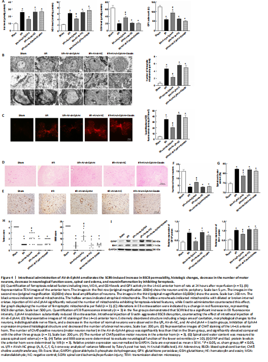
To evaluate whether EphA4 was involved in the regulation of SCIRI related-ferroptosis, we performed a rescue experiment using Erastin. First of all, we examined the effect of knocking down EphA4 on ferroptosis-related factors. The iron level and MDA content were significantly lower, and the GSH and GPX activities were remarkably higher, in theAV-sh-EphA4 group compared with the I/R and AV-sh-NC groups. Administration of Erastin reversed these effects (Figure 4A). Furthermore, we employed transmission electron microscopy to assess neuronal structure in the anterior horn and found that SCIRI induced neuronal damage. Injured neurons were irregular in shape and exhibited complete or local rupture of the cell membrane. The nuclear membranes were intact, locally sunken, wrinkled, or serrated. The mitochondria were shrunken, the internal cristae were dilated and broken, and the membrane density was increased, all of which are characteristic morphologic features of ferroptotic mitochondria. Mitochondria with ferroptosis-related morphological features also appeared in the axons. Injection of AV-sh-EphA4 markedly reduced the number of mitochondria exhibiting ferroptosis-related features, while Erastin administration reversed this effect (Figure 4B). The degree of damage to the BSCB was assessed by detecting EB leakage into the spinal cord tissue. The EB fluorescence intensity increased markedly after SCIRI, whereas inhibiting EphA4 expression substantially reduced EB extravasation. Moreover, intrathecal injection of Erastin counteracted the protective effect that intrathecal injection of AV-sh-EphA4 had on the BSCB (Figure 4C). Hematoxylin and eosin staining showed that the structure of the anterior horn was extremely disordered after SCIRI, the morphology of some neurons was irregular, and multipolarity disappeared. Neurons varied in their appearance, being shrunken, blunt, lightly stained, or diffusely swollen with eosinophilic cytoplasm, and the nuclei were blurred or pyknotic. In some tissue slices, a large area of cavitation was evident. Individual nerve fibers could not be distinguished. In the sham group, the neurons in the anterior horn were intact and exhibited fine, granular cytoplasm and Nissl substance. SCIRI decreased the number of intact neurons (Figure 4D). Immunohistochemical staining showed that the number of motor neurons in the anterior horn decreased markedly after SCIRI. In the AV-sh-EphA4 group, the number of motor neurons was markedly higher than that in the I/R group and AV-sh-NC group. Intrathecal injection of Erastin reversed the increase in motor neuron numbers in the anterior horn induced by intrathecal injection of AV-sh-EphA4 (Figure 4E and F). The degree of spinal cord edema showed a similar trend (Figure 4G). Next, we used Tarlov and BBB scores to assess the motor function of the lower extremities. Compared with the sham group, SCIRI substantially decreased motor function scores, while the AV-sh-EphA4 group showed markedly higher scores than the I/R and AV-sh-NC groups. Rats injected with AV-sh-EphA4 and Erastin had lower scores than rats injected with AV-sh-EphA4 alone (Figure 4H). Next, we performed western blotting for astrocyte and microglia markers to investigate the degree of proliferation and inflammatory state. SCIRI induced substantial increases in GFAP and Iba1 protein expression in the anterior horn. Inhibiting EphA4 expression markedly reduced GFAP and Iba1 expression, and Erastin reversed this effect. Taken together, these findings suggest that knocking down EphA4 suppresses inflammation by inhibiting ferroptosis (Figure 4I).
Figure 7|Immunofluorescence double-labeling of EphA4 and p-Erk1/2 or p-Beclin1 in the rat L4–L6 spinal cord anterior horn.
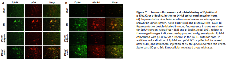
Fluorescence double-labeling showed that EphA4 co-localized with p-Beclin1 and p-Erk1/2, and that this co-localization was increased by SCIRI. However, inhibiting EphA4 expression decreased co-labeling. These finding support the Co-IP and western blotting results (Figure 7A and B).