脑损伤
-
Figure 1|Animal model establishment and laser speckle imaging of the brain.
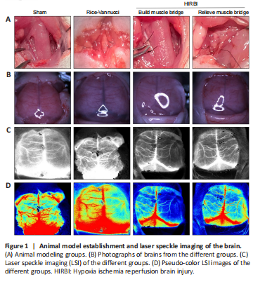
Sham group: The right CCA was exposed, and no other operations were performed. Rice-Vannucci group: The proximal and telecentric ends of the right CCA were ligated, and the artery between the ligation knots was cut carefully. After surgery, the rats were allowed to rest in a 37°C chamber for 10 minutes, and then they were subjected to hypoxia (8% oxygen and 92% nitrogen) in a hypoxia chamber for 120 minutes. HIRBI group: Briefly, the blood flow of the right CCA was blocked for 150 minutes (including 120 minutes of exposure to hypoxic circumstances), and then the right CCA muscle bridge was relieved by muscle bridge relief operation (Figure 1). The rats in each group were returned to their mothers after the operations.
The CCA muscle bridge technique can be used to construct a HIRBI model CBF status was assessed by examining physical images (Figure 1B), speckle images (Figure 1C), and pseudo-color images (Figure 1D) obtained by LSI. The LSI results showed that the left and right CBF were normal in the Sham group, whereas the right CBF was significantly decreased in the Rice-Vannucci and HIRBI groups. After the muscle bridge was removed, right CBF was restored.
Figure 6|Transmission electron microscopy (TEM) analysis of mitochondrial structures in the hippocampal CA1 region.
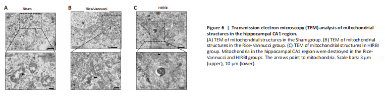
Due to the fact that most of the differentially expressed proteins were located in the mitochondria, we observed the mitochondria by TEM. In the Sham group, the mitochondrial membrane was intact, and the mitochondrial size and mitochondrial cristae apparatus were relatively normal (Figure 6A). Mitochondrial shrinkage was found in both the Rice-Vannucci and HIRBI groups. In addition, the mitochondrial membrane was ruptured, and the mitochondrial cristae were reduced or absent (Figure 6B and C); these features are consistent with ferroptosis (Chen et al., 2021; Lin et al., 2022). There was no discernible difference in degree of damage between the HIRBI and Rice-Vannucci models.
Figure 7|Immunofluorescence staining and Western blot analysis of the different groups.
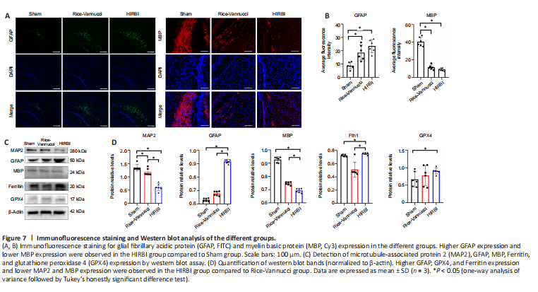
Based on the proteomic bioinformatics analysis results, we analyzed GFAP and MBP expression in the brain. GFAP and MBP are the most reliable and potential biomarkers for early assessment of hypoxic-ischemic brain injury and prediction of long-term neurological dysfunction in HIE neonates (Garcia-Alix et al., 1994; Lv et al., 2015). The level of GFAP expression in both the HIRBI and Rice-Vannucci groups was higher than that in the Sham group (P < 0.05; Figure 7A). However, the highest level of MBP expression was seen in the Sham group (P < 0.05; Figure 7B).