视神经损伤
-
Figure 1|Cell culture of MSCs from rat umbilical cord and collection of EVs.
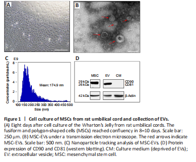
The growth of UC-MSCs was observed in the UC fragments several hours after incubation. The fusiform and polygon-shaped cells proliferated within 24 hours and reached confluency in 8 days (Figure 1A). Approximately 2.5 mL MSC-EVs were obtained from 50 mL MSC supernatant at a concentration of 1510.6 μg/mL determined by the bicinchoninic acid protein quantification kit (Beyotime). The cup-shaped UC-MSC-derived EVs were visualized under electron microscopy (Figure 1B). MSC-EVs were analyzed using nanoparticle tracking analysis; their concentration was 3.66 × 10¹¹ particles/mL with a mean diameter of 174.9 ± 21 nm (Figure 1C). The western blot assay revealed a specific positive expression of CD90 on UC-MSC and CD81 on EVs (Figure 1D).
Figure 3|Protective effect of umbilical cord-derived MSC-EV treatment on retina damage and ganglion cell loss induced by COH.
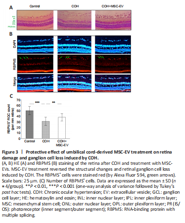
Compared to the normal rats, changes in the HE staining of the retina were observed in the COH rats, including marked swelling of the ganglion cell layer, the inner plexiform layer, the outer nuclear layer, and the RGC layer of the retina. In addition, in the RGC layer, the number of cells was reduced, with irregularity in the cell arrangement, darkened staining, and condensation of the cell nuclei. In the MSC-EV treatment group, the swelling of the retinal layers was alleviated, and the morphological anomaly was improved in the RGC layer (n = 3/group; Figure 3A).
When stained with the RGC-specific marker RBPMS (Kwong et al., 2010; Rodriguez et al., 2014), the number of RGCs decreased significantly (P < 0.001, n = 4/group, Figure 3B and C) in the COH eyes. With the treatment of MSC-EV, this decrease in RGC number was significantly smaller than that of the COH eyes (P < 0.01, n = 4/group).
Figure 4|Inhibition of apoptosis by MSC-EV treatment in the COH rat retina.
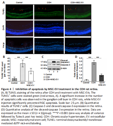
TUNEL staining of the retina revealed a significant increase in cell apoptosis in rats with COH (P < 0.001, n = 3, vs. control group). After treated with MSC-EV injection, the number of apoptotic cells significantly decreased in the COH rats (P < 0.001, n = 3, vs. COH group; Figure 4A and B).
The Western blot assay revealed that the expression of cleaved caspase-3, which is an apoptosis-related protein (Julien and Wells, 2017; Asadi et al., 2022) was significantly up-regulated in the retina of COH rats (P < 0.001, n = 3/group, vs. control group). After MSC-EV treatment, cleaved caspase-3 expression was significantly inhibited (P < 0.001, n = 3/group, vs. COH group; Figure 4C and D).
Figure 5|Neuroprotective effects of MSC-EV treatment on RGCs in the COH rat retina.
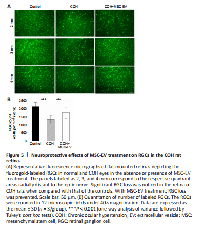
Representative images of RGC fluorogold staining are shown in Figure 5A. Quantitative calculation revealed that fluorogold-labeled RGC in the retinas of COH rats decreased significantly at week 8 (P < 0.001, n = 3/group, vs. control group). For the COH eyes that received MSC-EV injection, the RGC density was significantly higher than those without the MSC-EV treatment (P < 0.001, n = 3/group, Figure 5B).