脊髓损伤
-
Figure 1|Liproxstatin-1 reduces blood-spinal cord barrier disruption after SCI.
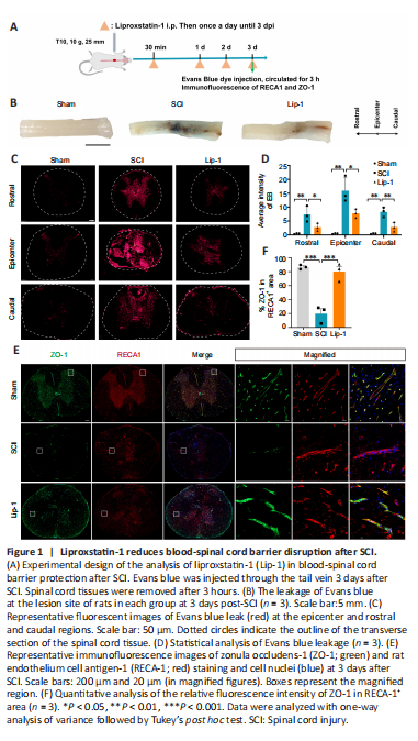
Disruption of the BSCB is a major cause of secondary injury in the acute phase of SCI. Thus, ameliorating BSCB injury is a feasible target for SCI repair (Lee et al., 2012; Zhang et al., 2017). We first examined whether Lip-1 influenced BSCB disruption in the acute phase of SCI (Figure 1A). We observed leakage of Evans Blue in the SCI group compared with the Sham group, indicating that the barrier function of BSCB was damaged. After Lip-1 treatment, the leakage of Evans Blue had sharply decreased (Figure 1B). Evaluation of fluorescence intensity of Evans Blue revealed that the SCI group exhibited significant Evans blue leakage at the epicenter (P < 0.01), as well as rostral (P < 0.01) and caudal (P < 0.01) spinal cord segments; Lip-1 reduced Evans blue leakage at all locations (P < 0.05, P < 0.05, P < 0.01 for epicenter, rostral and caudal areas, respectively; Figure 1C and D).
Paracellular tight-junction proteins (TJP) such as zonula occludens-1 (ZO-1) are responsible for maintaining the integrity of the BSCB (Zhao et al., 2022). We next examined the effect of Lip-1 on TJP ZO-1 expression in ECs by immunofluorescence staining. ZO-1 expression in ECs was downregulated after injury and this was reversed by Lip-1 administration (Sham vs. SCI, P < 0.001; SCI vs. Lip-1, P < 0.001; Figure 1E and F). Taken together, these results indicate that Lip-1 attenuates BSCB disruption by upregulating ZO-1 expression in ECs.
Figure 3|Liproxstatin-1 inhibits bEnd.3 cell ferroptosis and increases tight junction protein expression.
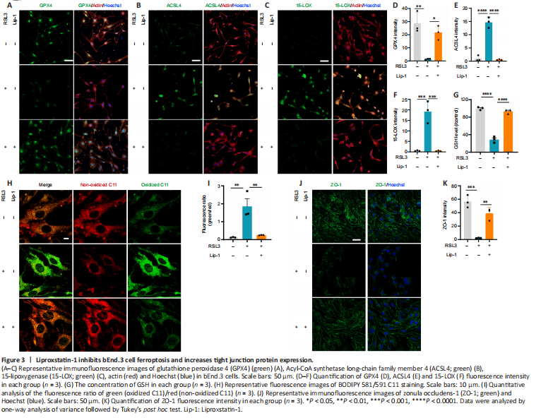
Immunocytochemistry showed that GPX4 expression in bEnd.3 cells was significantly decreased after RSL3 treatment (P < 0.01) and rescued by Lip-1 treatment (P < 0.05; Figure 3A and D). Ferroptosis markers ACSL4 and 15-LOX were elevated in the RSL3-treated group (ACSL4, P < 0.0001; 15-LOX, P < 0.001), indicating a high level of lipid peroxidation in ferroptotic bEnd.3 cells. After Lip-1 treatment, the two markers were reduced (ACSL4, P < 0.0001; 15-LOX, P < 0.001; Figure 3B, C, E, F). The GSH content was next evaluated; compared with RSL3 treatment alone, Lip-1 significantly increased GSH concentration (P < 0.0001; Figure 3G). The fluorescent ratio of oxidized BODIPY C11/non-oxidized C11 in RSL3-treated bEnd.3 cells was significantly increased compared with ratios in RSL3 and Lip-1 co-treated bEnd.3 cells (P < 0.01; Figure 3H and I). This indicates a decrease of lipid peroxidation in bEnd.3 cells after Lip-1 treatment. RSL3 significantly decreased expression of the tight junction protein ZO-1, whereas Lip-1 significantly increased the expression of ZO-1 (P < 0.01; Figure 3J and K). Taken together, these results indicate that Lip-1 suppressed RSL3-induced bEnd.3 cell ferroptosis and upregulated the expression of ZO-1.
Figure 4|Liproxstatin-1 regulates ferroptosis of vascular endothelial cells after SCI.
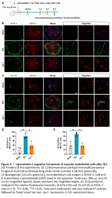
We next examined whether ECs in rats underwent ferroptosis after SCI (Figure 4A). We used RECA1 as marker of vascular ECs (Betterton et al., 2022). ACSL4 and 15-LOX were not detected in ECs of the Sham group, whereas their expression increased significantly after SCI (ACSL4, P < 0.01; 15-LOX, P < 0.01; Figure 4B–E). Notably, ECs in the Lip-1 group showed significantly decreased expression of ACSL4 and 15-LOX (ACSL4, P < 0.05; 15-LOX, P < 0.01; Figure 4B–E). These results indicate that in acute SCI, ECs undergo ferroptosis, and Lip-1 inhibition of ferroptosis is associated with decreased expression of ACSL4 and 15-LOX.
Figure 5|Liproxstatin-1 inhibits the infiltration of macrophages and neutrophils after SCI.
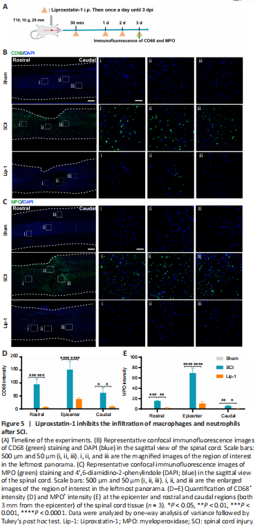
Upon destruction of the BSCB after SCI, blood-derived cells such as macrophages and neutrophils infiltrate into the spinal cord parenchyma to aggravate the inflammatory cascade (Sharma, 2011; Jin et al., 2021). Activated M1 macrophages release a large number of pro-inflammatory factors, and neutrophils infiltrating the spinal cord release MPO, catalyzing an inflammatory response (Beck et al., 2010; Lee et al., 2011; Neirinckx et al., 2014). Therefore, we evaluated the effect of Lip-1 treatment on inflammatory cells by immunofluorescence 3 days after SCI (Figure 5A). Many CD68+ macrophages (rostral, P < 0.001; epicenter, P < 0.0001; caudal, P < 0.05; Figure 5B and D) and MPO+ neutrophils (rostral, P < 0.001; epicenter, P < 0.0001; caudal, P < 0.01; Figure 5C and E) significantly infiltrated into the spinal cord in the SCI group compared with the Sham group. However, Lip-1 significantly attenuated the increased infiltration of inflammatory cells at the epicenter (CD68, P < 0.0001; MPO, P < 0.0001), rostral (CD68, P < 0.001; P < 0.01), and caudal (CD68, P < 0.05; MPO, P < 0.05). This suggests that Lip-1 attenuates the disruption of the BSCB, thus reducing the infiltration of peripheral-derived inflammatory cells into the spinal cord parenchyma.
Figure 6|Liproxstatin-1 alleviates astrogliosis after SCI.
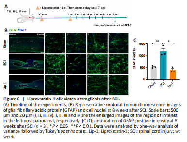
Astrocytes are excessively activated following SCI, forming glial scars that hinder axon growth. To evaluate the effect of Lip-1 on astrocytes after SCI, immunofluorescence staining of the astrocyte marker glial fibrillary acidic protein (GFAP) was performed (Figure 6A). Eight weeks after SCI, there was obvious astrogliosis around the epicenter; Lip-1 significantly reduced the astrogliosis (P < 0.05; Figure 6B and C). Moveover, Lip-1 reduced the infiltration of peripheral inflammatory cells into the spinal cord parenchyma to improve the inflammatory microenvironment at the injury site, which is a more favorable environment for neuronal survival. Therefore, we further examined the protective effect of Lip-1 on neurons by immunofluorescence staining at 8 weeks after SCI (Additional Figure 3A). We found that Lip-1 intervention effectively inhibited the loss of neurons around the lesion (P < 0.01; Additional Figure 3B and C). We conclude that Lip-1 increased neuronal survival and reduced the glia scars after SCI.