神经退行性病
-
Figure 1|Progressive amyloid-β pathology and cognitive dysfunctions in 5×FAD mice.
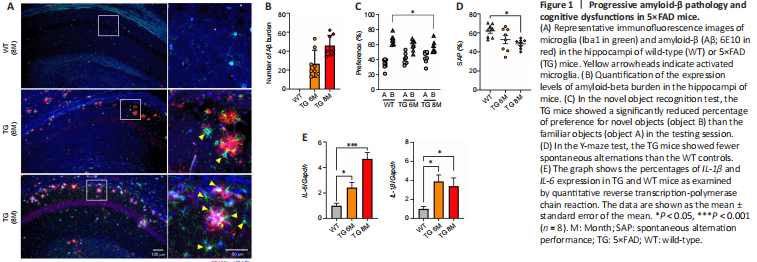
5×FAD, a transgenic AD mouse model, exhibits many AD-related phenotypes and a relatively progressive and aggressive Aβ production. Immunofluorescent staining for Aβ showed that the number of Aβ plaques increased in TG mice with aging, as expected (Figure 1A and B). Immunostaining for the microglia marker, Iba1, showed increases in microglial densities and activated morphology at 6 and 8 months of age in the hippocampus of 5×FAD mice compared with WT mice at the age of 8 months. Activated microglia were more densely clustered around core plaque with aging in 5×FAD mice.
To examine cognitive functions in TG and WT mice, we performed the novel object recognition memory and Y-maze tests in WT and TG mice at the age of 6 and 8 months. At the age of 8 months old, the TG mice showed more severe cognitive impairments in preferential recognition of the novel object (Figure 1C) and reduced spontaneous alternation in the Y-maze test as well when compared with those of 6 months old, indicating that 5×FAD mice showed age-dependent behavioral deficits (Figure 1D). In addition, we analyzed the mRNA expression of genes encoding proinflammatory cytokines, including IL-1β and IL-6, in the mouse hippocampus. Compared to those in WT mice, the expression levels of IL-1β and IL-6 were significantly increased in the TG mouse hippocampus (Figure 1E; P < 0.05). These observations showed that cognitive dysfunctions in 5×FAD mice were progressive and worsen with age, which was accompanied by increases in levels of inflammation-related genes in the hippocampus.
Figure 3|Effects of RF-EMFs on the levels of Aβ and APP processing in the hippocampi of mice.
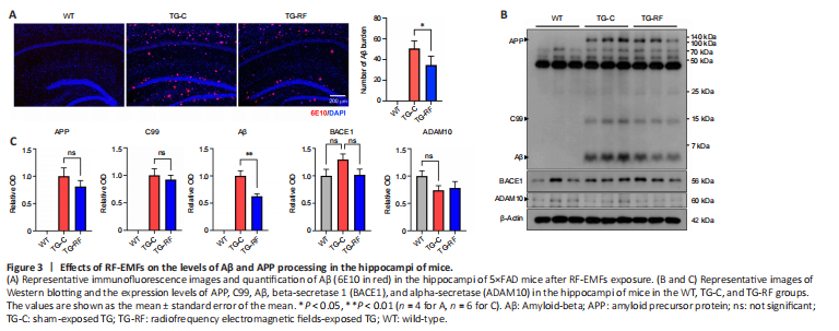
To demonstrate the effects of RF-EMFs on Aβ accumulation, we first examined the levels of Aβ deposition by using 6E10 staining. Immunofluorescence analysis revealed decreases in Aβ plaque deposition in the hippocampi of the TG-RF group compared with the TG group (Figure 3A). To further examine the effects of RF-EMFs on APP processing, Western blotting was performed to analyze the protein expression in the hippocampi of 5×FAD mice (Figure 3B). The APP, C99, and Aβ levels were increased significantly in TG mice compared with the age-matched WT mice. Although the APP and C99 protein levels were not significantly altered by RF-EMFs exposure, the Aβ expression in 5×FAD mice exposed to RF-EMFs was significantly decreased compared with that in TG mice (Figure 3C). Apart from that, the BACE1 and ADAM10 levels did not differ between TG mice and those of the WT and TG-RF groups (Figure 3C). These results suggest that RF-EMFs exposure is sufficient to decrease the production and deposition of Aβ.
Figure 5|Effects of radiofrequency electromagnetic fields on Iba1 and CSF1R in the hippocampi of 5×FAD mice.
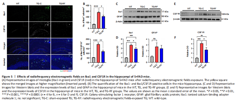
To determine whether RF-EMFs exposure prevents the activation of microglia, we examined the levels of Iba1 (a microglial marker) and CSF1R (a microglia proliferative marker) by using specific markers. The number of Iba1-positive and Iba1/CSF1R-double positive cells in the hippocampi of each group were counted (Figure 5A and B). In the TG group, there are significant increases in Iba1- and Iba1/CSF1R-expressed microglia compared to the WT group. As known that CSF1R is expressed in microglia (Hagan et al., 2020; Han et al., 2022), CSF1R expression is overlapped in most of Iba1-positive microglia as seen in the inserted image of Figure 5A. The number of microglia expressing CSF1R remarkably reduced in the hippocampus of the TG-RF group. The expression levels of Iba1 and GFAP, a biomarker of astrocytes, were increased in the hippocampal region of TG mice compared with WT mice as determined by Western blotting (Figure 5C and D); RF-EMFs significantly reduced the expression of Iba1 in the TG-RF group compared with the sham-exposed TG group. However, the difference in the GFAP levels did not reach statistical significance (Figure 5C and D).
Next, we investigated whether the expression of CSF1R was altered by RF-EMFs exposure. Western blot analysis revealed significant increases in the levels of CSF1R in the TG mouse hippocampus compared with WT mice (P < 0.001; Figure 5E and F). However, these increases were significantly ameliorated in the TG-RF group relative to the TG group (P < 0.05; Figure 5E and F), which was consistent with the expression of Iba1, suggesting that neuroinflammation was ameliorated by RF-EMFs exposure in the AD mouse model.