周围神经损伤
-
Figure 5|Histological changes in the biceps brachii.
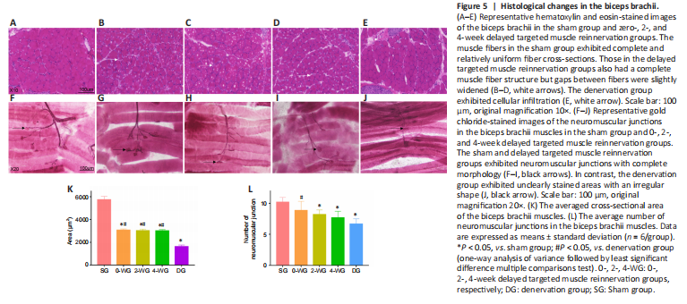
Myofiber morphology in the biceps brachii muscles
Muscle fibers in the sham group were tightly packed and relatively uniform (Figure 5A). Those in the dTMR groups had a similar morphology with complete structure but the gaps between fibers were slightly widened (Figure 5B–D). In the denervation group, fibrosis and cellular infiltration were present between individual fibers; gaps between fibers were also widened (Figure 5E). Muscle fiber CSA was significantly higher in the sham group than the other groups (P < 0.05; Figure 5K). CSA did not significantly differ among the dTMR groups (P > 0.05). CSA was significantly higher in the dTMR group than the denervation group (P < 0.05).
NMJs in the biceps brachii muscles
The NMJs in the sham and zero-dTMR groups were complete and exhibited a clear color and oval shape (Figure 5F and G). Intact shape of NMJs can be found in the 2- and 4-week dTMR groups (Figure 5H and I). In the denervation group, there were unclearly stained areas with an irregular shape (Figure 5J). As shown in Figure 5L, the number of NMJs did not significantly differ between the sham and zero-week groups (P > 0.05). The number of NMJs was lower in the 2- and 4-week dTMR and denervation groups than the sham group (P < 0.05). The number of NMJs did not significantly differ among the three dTMR groups (P > 0.05). Compared with the sham and zero-week dTMR groups, the number of NMJ in the denervation group was significantly lower (P < 0.05).