周围神经损伤
-
Figure 2|PF4 is expressed in rat sciatic nerves and Schwann cells.
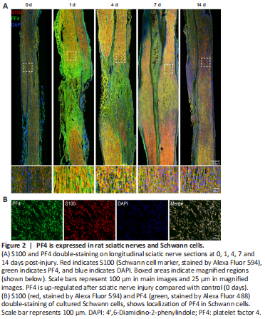
Immunostaining of longitudinal sciatic nerve sections showed that PF4 protein expression was also elevated after sciatic nerve crush injury (Figure 2A). Moreover, up-regulated PF4 localized with S100 signal, a marker of Schwann cells (Figure 2A). In cultured primary Schwann cells, immunostaining with anti-PF4 and anti-S100 antibodies further demonstrated co-localization of PF4 and S100, indicating the presence of PF4 in Schwann cells (Figure 2B).
Figure 3|PF4 silencing inhibits Schwann cell viability, proliferation, and migration.
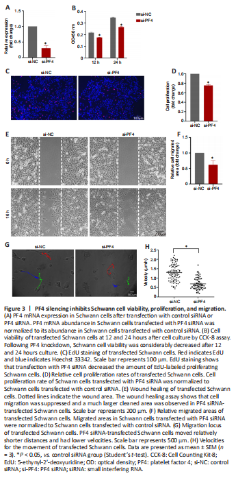
Cultured primary Schwann cells were transfected with a siRNA against PF4 to evaluate the functional roles of PF4. PF4 siRNA had a high knockdown efficiency and successfully reduced PF4 mRNA abundance to about 30% (Figure 3A). Following PF4 knockdown, Schwann cell viability was considerably reduced after 12-hours of culture (Figure 3B). By 24 hours, the viability of both PF4 siRNA-transfected and control siRNA-transfected Schwann cells were elevated to higher levels. Yet Schwann cells transfected with PF4 siRNA were less viable than cells transfected with control siRNA (Figure 3B). EdU staining showed that transfection with PF4 siRNA decreased the amount of EdU-labeled proliferating Schwann cells (Figure 3C). Summary data showed that PF4 knockdown reduced the proliferation rate of Schwann cells to less than 80% (Figure 3D). In addition to cell viability and proliferation, we examined the movement of Schwann cells using a wound healing assay. This demonstrated that cell migration was suppressed and a much larger cleaned area was observed in PF4 siRNA-transfected Schwann cells after removal of the wound healing culture insert (Figure 3E and F). Using a live-cell imaging system (He et al., 2018), Schwann cell movement was directly visualized. The migration locus of representative Schwann cells was marked (Figure 3G). Whereas PF4 siRNA-transfected Schwann cells had relatively shorter movement distances and lower velocities (Figure 3H). These observations indicate that PF4 silencing suppresses Schwann cell viability, proliferation, and migration.
Figure 4|PF4 recombinant protein supports Schwann cell proliferation and migration. 、
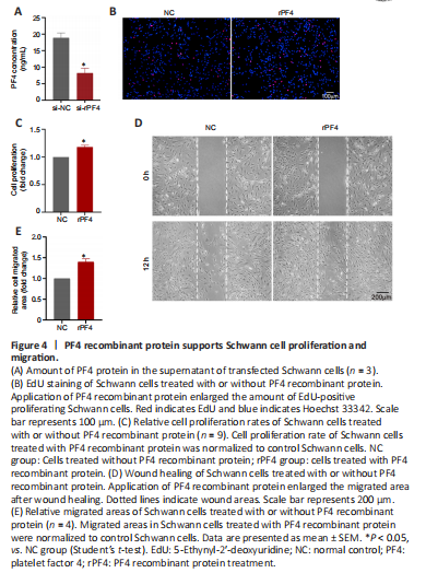
Results from ELISA showed that PF4 protein could be detected in the supernatant of cultured Schwann cells and the amount of secreted PF4 protein was decreased after PF4 siRNA transfection (Figure 4A). To mimic the effects of secreted PF4 protein, Schwann cells were exposed to PF4 recombinant protein. In contrast to PF4 knockdown, application of PF4 recombinant protein enlarged the amount of EdU-positive proliferating Schwann cells (Figure 4B and C) and the migrated area after wound healing (Figure 4D and E). Together, this supports a role of PF4 in promoting Schwann cell proliferation and migration.
Figure 5|PF4 recombinant protein supports axon growth and elongation.
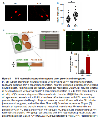
PF4 recombinant protein was further applied to cultured neurons to investigate the effects of PF4 on neuronal behavior. Following addition of PF4 recombinant protein, neurons had a visibly increased neurite length (Figure 5A and B), demonstrating that PF4 encourages axon growth. Next, neurons were cultured in microfluidic chambers (Figure 5C). Axons were exposed to injuries and subsequent regeneration conditions of damaged axons were examined. After 48-hours of culture, axons were found to regenerate to a certain length. However, after treatment with PF4 recombinant protein, the regenerated length of injured axons increased by about 5-fold (Figure 5D and E). These observations show that PF4 protein accelerates the growth and regeneration of axons.
Figure 6|PF4 silencing inhibits Schwann cell proliferation following sciatic nerve injury.
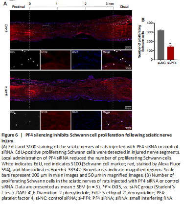
The in vivo effect of PF4 was preliminarily studied by delivering PF4 siRNA into adult Sprague-Dawley rats. PF4 siRNA was injected into injured sciatic nerves immediately after nerve crush injury. One day after nerve injury and PF4 siRNA injection, rat sciatic nerves were collected and immunostained with EdU and S100 to visualize Schwann cell proliferation. EdU-positive proliferating Schwann cells were detected in injured nerve segments (Figure 6A). Quantification of the number of proliferating Schwann cells in the sciatic nerves of rats injected with PF4 siRNA or control siRNA demonstrated that local administration of PF4 siRNA reduced the amount of proliferating Schwann cells (Figure 6A and B). This indicates that PF4 silencing suppresses Schwann cell proliferation following peripheral traumatic nerve injury.