脑损伤
-
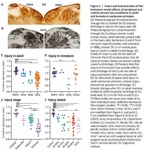
Figure 2|Intact and lesioned sides of the brainstem reveal effects of peripheral and central sensory loss conditions in adult and immature nonhuman primates.

结果:For decades, researchers have known that when neurons lose their activating inputs, they can atrophy and even die. For example, removing sensory nerve inputs from the arm to the spinal cord after amputation or cutting the spinal nerves can shrink the post-synaptic target zones (e.g., Woods et al., 2000). From touch receptors for the hand and arm, primary nerve afferents enter the spinal cord, and axons travel in the dorsal columns to target the cuneate nucleus (Cu) of the brainstem on the same side of the body (Figure 1). When sensory loss is unilateral, the overwhelming result is for the associated primary target zone to shrink in total size (cross-sectional area), as compared to the size of the opposite side. While this concept is widely accepted, many details about the factors that influence the shrinkage are not known. What are the effects of different types of sensory loss on the relative shrinkage? Are the effects different whether the sensory loss occurred during development or adulthood? What are the implications of size reduction due to sensory loss? Qi et al. (2023) examined the histological consequences of sensory loss of the hand and arm on the Cu in nonhuman primates (Figure 2A and B). Historically over 40 years, Kaas and colleagues reported on the plasticity in the cortex of nonhuman primates that experienced experimental and therapeutic unilateral sensory loss (for specific references, see Qi et al., 2023). In many of the same cases that were studied for cortical effects, tissue sections of the brainstem were histologically processed for cytochrome oxidase, revealing cytoarchitecture of the Cu in 122 cases, which included 37 Old World macaque monkeys. The remaining 85 cases included prosimian Galagos and New World monkeys. The sizes of left and right Cu were measured for each brainstem section in the histological series, and the ratio of the sizes in cases with and without unilateral sensory loss was calculated. Types of sensory loss studied included the transection of one or more peripheral sensory nerves with or without manipulations of regeneration, therapeutic amputations ranging from digits to the forelimb, and central spinal cord injury of the primary somatosensory dorsal column pathway.
Sensory loss effects in the brainstem cuneate nucleus of immature versus adult nonhuman primates: Across all types of sensory loss studied by Qi et al. (2023), the sex or species identity had no significant effect on the ratio of the left and right Cu size. With an evolutionary perspective in mind, if the effects of sensory loss are similar across different species, they are likely relevant to the effects in humans. The influence of the type of sensory loss on the ratio of the Cu size was examined separately for cases that had unilateral sensory loss events in immaturity and cases that experienced sensory loss in adulthood (Figure 2C and D). Overall, there was a tendency for sensory loss to cause the deprived Cu to shrink significantly, except in immature subjects where peripheral nerve damage caused no detectable shrinkage. The detected effects on the size of the Cu were those related to the severity or type of sensory loss, such that single nerve cut results in limited shrinkage versus the rare cases of therapeutic amputation of an injured arm. The results from Qi et al. (2023) general linear regression analyses illustrate the expected principle that greater Cu shrinkage is related to a more severe extent of sensory loss. They also indicate that in developing primates, the Cu does not shrink after most peripheral nerve injuries, and significant shrinkage in immature subjects only occurred in the few (3) cases of massive sensory loss caused by arm amputation.
Similar effects of central and peripheral sensory loss: Unilateral dorsal column lesions of the spinal cord were performed in adult cases only, and the lifetime living with sensory loss (recovery time span between the injury and histological processing) and the severity of the dorsal column loss were factors considered for their influences on the Cu size ratios (Figure 2E and F). The time span of dorsal column injury was divided into subacute (48 hours to 14 days), intermediate (14 days to 6 months), and chronic (> 6 months) stages (Ahuja and Fehlings, 2016, review). Following spinal cord injury, longer lifetime living with sensory loss was associated with reduced Cu size (Figure 2E). For adult animals with spinal cord injury, the proportion of the dorsal column pathway damaged was related to the size reduction ratio of the Cu (Figure 2F), similar to the relationship of peripheral sensory loss to the size of the Cu. Grouping peripheral and central sensory loss types to examine the effects of different sensory loss characteristics, there was a significant interaction between the extent of sensory loss and lifetime living with sensory loss (“recovery time span”), such that longer recovery time span strongly related to greater Cu shrinkage in cases after spinal cord injury, followed by cases after amputation. Thus, sensory loss at peripheral or central levels appears to affect Cu shrinkage in similar ways relative to sensory loss severity.
While not directly shown by Qi et al. (2023), we have found that for any type of adult-onset sensory loss studied to date, both peripheral and central types, when the Cu shrinks, the cell bodies tend to become more densely packed rather than sparse, such as seen when tissue is processed to reveal neuron nuclei with the marker NeuN (Figure 2B). Neuron atrophy in the spinal cord and Cu (including shrinking of the cytoplasm of cell bodies and neurites and retraction of synaptic arborizations) is the likely cause of Cu volume reduction in response to sensory loss. Such neuron atrophy is known to occur when axons are cut or damaged (e.g., Woods et al., 2000). The densely packed neuron nuclei in the Cu suggest that atrophy of the neurons and neuropil in the Cu does not necessarily result in neuron death, similar to the findings of Woods et al. (2000). After primary sources of input are lost, secondary and modulating sources of inputs can maintain the neurons, even though neuron cell bodies and their processes shrink. These surviving neurons may be key targets for therapeutic strategies after sensory loss.
点击此处查看全文
Implications for brainstem recovery from studies in primates after sensory loss from arm
发布日期: 2023-09-02 浏览:
83