神经损伤与修复
-
Figure 1|Ultrastructural features of the endoplasmic reticulum in primary microglia in culture.
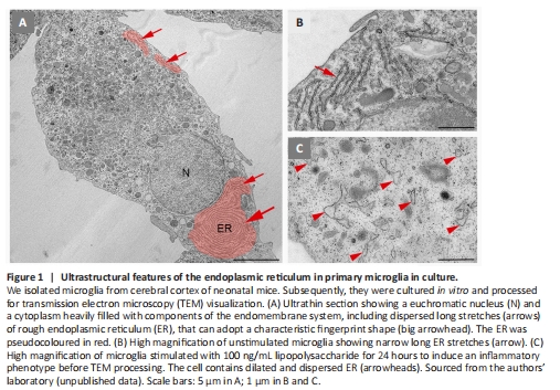
The ER constitutes a large and dynamic compartment that forms a continuous network of sheet-like cisternae with ribosomes on the cytosolic face of the membrane, the rough ER, and tubules lacking ribosomes, the smooth ER. Both differ in physical and functional characteristics. In microglial cells, the content of the rough ER is very abundant, and its ultrastructure can be easily visualized by TEM in ultrathin sections of brain tissue (Savage et al., 2018) or of primary microglial cell cultures (Figure 1). Cisternae may appear as long stretches arranged in parallel stacks or form extensive concentric systems resembling fingerprints. The lumen of the cisternae is quite narrow, but it might appear dilated by elevated production and secretion of cytokines and other factors, especially in the inflammatory microglial phenotype (Figure 1C). We can also find dilated ER under stress or pathological conditions (Bisht et al., 2016). Functionally, it is well known that the ER is involved in the synthesis, folding, modification, and transport of proteins and lipid metabolism, but the ER also represents the largest intracellular compartment for Ca2+ storage in all cells, including microglia. However, many questions remain unanswered about how this reservoir can participate in modulating Ca2+ signals in response to extra- or intracellular cues, microglial activation states, lesions, or pathologies.