脑损伤
-
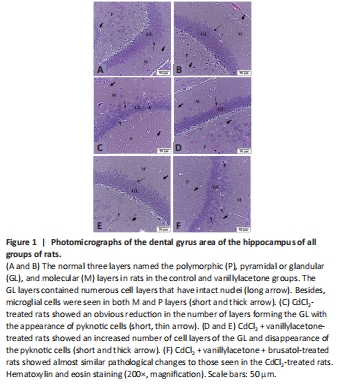
Figure 1|Photomicrographs of the dental gyrus area of the hippocampus of all groups of rats.

Typical dental gyrus areas of the hippocampi, characterized by intact three layers [i.e., the polymorphic (P), pyramidal or glandular (GL), and molecular layers], were detected in both the control and vanillylacetone-treated rats (Figure 1A and B). In both groups, abundant microglial cells with multiple layers forming the GL were seen. In addition, few glial cells were seen in both groups of rats’ P and M layers (Figure 1A and B). An increased number of microglial cells in both the P and M layers, with an evident decrease in the number of layers forming the GL layers, was observed in the CdCl2-treated rats, and many pyknotic cells were also seen in this group of rats (Figure 1C). The number of layers and cells forming the GL and microglial cells in the P and M layers were significantly reduced in rats treated by CdCl2 and vanillylacetone, with a reduction in the number of pyknotic cells in the GL (Figure 1D and E). However, inhibition of Nrf2 by brusatol (CdCl2 + vanillylacetone + brusatol) induced similar pathological changes in the hippocampi observed in the model group (CdCl2-treated rats; Figure 1F).