视神经损伤
-
Figure 2 | Effect of simulated weightlessness on retinal morphology in rats.
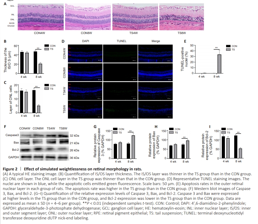
To determine whether simulated weightlessness can cause retinal damage, HE and TUNEL staining were performed to assess alterations in retinal structure and detect apoptosis, respectively. HE staining showed that, compared with the CON group, the IS/OS thickness (indicating optic cone and rod cell numbers) and the outer nuclear layer thickness were lower in the TS8W group (P < 0.05; Figure 2A–C). Furthermore, TUNEL staining showed a significant number of apoptotic cells in the outer nuclear layer after 8 weeks of tail suspension (P < 0.01; Figure 2D and E). Consistent with these findings, higher expression levels of the proapoptotic molecules Bax and Caspase 3 were detected in the TS8W group than in the CON group (Figure 2F–I). This suggests that tail suspension simulation of weightlessness can lead to degenerative changes in apoptosis of the outer nuclear layer cells of the rat retina.
Figure 4 | Effect of simulated weightlessness on Müller cell reverse differentiation.
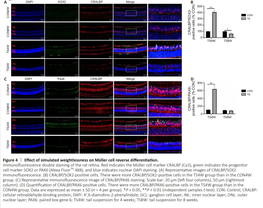
To determine whether simulated weightlessness stimulates retrodifferentiation of Müller cells, the retinas were double stained for CRALBP (a marker of Müller glial cells (Jurkute and Robson, 2021)) and SOX2 or PAX6 (stem/progenitor cell markers (Lin et al., 2009)). As shown in Figure 4A–D, following 4 weeks of tail suspension, the rats exhibited massive activation of retinal Müller cells and a statistical increase in SOX2 and PAX6 expression compared with the CON group (P < 0.01; Figure 4B and D). Following 8 weeks of tail suspension, a notable reduction was observed in the expression levels of SOX2 and PAX6, with SOX2 expression more greatly reduced (P < 0.05; Figure 4B). This suggests that tail suspension in rats effectively simulates weightlessness and stimulates retrodifferentiation of retinal Müller cells, which exhibit progenitor cell properties for a short period of time.
Figure 5 | Effect of simulated weightlessness on Müller cell neural differentiation.
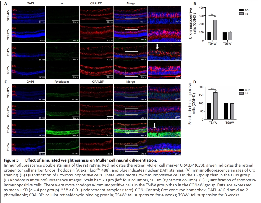
To determine whether simulated weightlessness stimulates Müller cell neurodifferentiation, we performedimmunofluorescence staining for crx and rhodopsin. Based on the results of proteomic analysis, we found that the expression of rhodopsin in the retina tissue of TS8W group rats decreased (Additional Table 1). Next, retinas were double stained for CRALBP and the photoprecursor/mature photoreceptor cell markers crx and rhodopsin. As shown in Figure 5, crx and rhodopsin expression levels were higher in the retinal tissues of rats in the TS4W group compared with the CON group (P < 0.01). This suggests that tail suspension simulates the early stage of weightlessness and promotes Müller cell neurodifferentiation.
Figure 6 | Simulated weightlessness can lead to Müller cell gelatinization.

To determine the effect of simulated weightlessness on gelatinization in retinal tissues, we detected the glial-associated marker GFAP and the glutamate (GLU) metabolism–related markers GS and GLAST in retinal tissues from rats in each group (Figure 6). Increased GFAP expression and decreased GS and GLAST expression were observed after 4 weeks of tail suspension compared with the CON group, although these differences were not statistically significant. GFAP expression was further increased (P < 0.01), and GS expression was further decreased (P < 0.01), in the retinal tissues of rats after 8 weeks of tail suspension compared with the CON group. GLAST is involved in the GLU cycle (Wunderlich et al., 2010), and significantly decreased GLAST expression was observed at 8 weeks of tail suspension (P < 0.01). Müller cells undergo a glial cell fate in the late stage of microgravity injury, which can affect GLU translocation and metabolism.