周围神经损伤
-
Figure 2 | PEG-fusion produces immediate restoration of electrophysiological continuity and preservation of many distal axons.
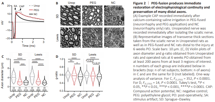
The status of axonal fusion/repair following a complete sciatic transection was confirmed using several well-published criteria (Bittner et al., 2016a; Ghergherehchi et al., 2016, 2019a, b, 2021; Mikesh et al., 2018a, b) as documented in Figure 2. Compound action potential recordings Figure 2A shows compound action potential (CAP) recordings of Unoperated sciatic nerve as well as PEG-fused and NC sciatic nerves immediately following calcium-containing saline application. A prominent CAP after the stimulus artifact was observed in PEG-fused rats but not in NC rats. This restoration of electrophysiological continuity strongly implies the restoration of axolemmal and axoplasmic continuity in some axons in PEG-fused sciatic nerves. Axon morphology Figure 2B illustrates axon morphologies in distal segments of sciatic nerves in Unoperated rats as well as PEG-fused and NC rats at 6 weeks PO. Unoperated rats exhibit similar (P > 0.05) axon diameter (SD: 4.14 ± 0.07 μm, Lewis: 4.34 ± 0.08 μm) and g-ratio (SD: 0.599 ± 0.003 μm, Lewis: 0.585 ± 0.003 μm) across strains (Figure 2C and D). Some statistical significance in g-ratio, but not axon diameter, was noted among the Unoperated subjects (data not shown), though such differences often even exist within the same animal at different locations (see Mikesh et al., 2018b for variation of axon diameter and g-ratio across proximal, graft, and distal portion of Unoperated rats). Most of the sampled operated rats were males (7 out of 8 cases; Table 1) to enable comparisons with historical data characterizing females(Mikesh et al., 2018a, b). NC distal nerves contained much debris and mostly small diameter (1–4 μm), thinly myelinated axons identified as regenerating axonal outgrowth in previous studies (Mikesh et al., 2018a, b). In contrast, PEG-fused distal nerves contained relatively little debris, some regenerative axons, but also some large diameter axons (4–7 μm) with thick myelination. These axons have been identified as successfully PEG-fused axons that do not undergo WD (Mikesh et al., 2018a, b). Overall, axons in PEG-fused nerves (SD: 2.34 ± 0.06 μm; Lewis: 2.44 ± 0.04 μm) had significantly larger diameters (P < 0.0001; Figure 2C) than those in NC nerves (SD: 1.89 ± 0.05 μm; Lewis: 1.80 ± 0.03 μm) in both strains. Axons in PEGfused nerves (SD: 0.580 ± 0.004; Lewis: 0.572 ± 0.003) had higher g-ratio than those in NC nerves (SD: 0.558 ± 0.007; Lewis: 0.561 ± 0.004) in both strains (Figure 2D), though there was significance in SD rats (P < 0.05) but not in Lewis rats (P = 0.13). No significant difference (P > 0.05) was found in axon diameter or g-ratio in the same treatment group across strains. Furthermore, because axon diameters and g-ratio of all groups in this study using mostly males were within the range previously reported for female SD rats (Mikesh et al., 2018a, b), our results suggested that sex difference in axon morphologies is also unlikely.