神经退行性病
-
Figure 1 | Exosome isolation and characterization.
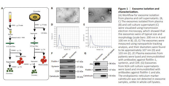
Exosomes were isolated from plasma and cell culture media (fetal bovine serum [FBS] without exosomes) as described previously (Lobb et al., 2015; Li et al., 2021). The workflow is shown in Figure 1A
The workflow for exosome isolation from the plasma samples and cell culture supernatants is shown in Figure 1A. TEM analysis showed that the isolated exosomes displayed a typical dish- or cup-like morphology and double lipid layers. Figure 1B shows the morphology of the exosomes isolated from plasma, and Figure 1C shows the morphology of the exosomes isolated from the cell culture supernatants. NTA showed that the average size of the exosomes isolated from plasma and cell culture supernatants were approximately 107 and 103 nm, respectively (Figure 1D and E). The plasma-derived exosomes expressed the exosome markers CD9, Syntenin, Alix, and Flotiliin-1, but not the negative control Calreticulin (Figure 1F). Similarly, the cell culture supernatant-derived exosomes expressed Alix and Flotiliin-1, but not Calreticulin (Figure 1G).
Figure 5 | NAGLU and α-syn expression in MPP+ -treated N2A cells and α-syn-overexpressing SH-SY5Y cells.
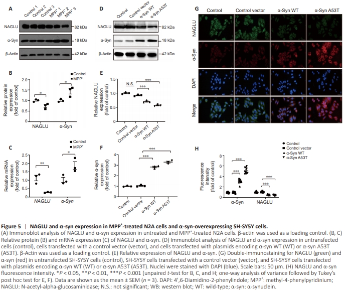
Eliminating aggregated α-syn is a potential therapeutic strategy for PD (Garrido et al., 2019). First, we assessed the NAGLU and α-syn expression in MPP+ -treated and untreated N2A cells by WB. The results showed that NAGLU expression was downregulated (P = 0.030), while α-syn expression was upregulated (P = 0.031), after MPP+ treatment (Figure 5A and B). Consistent with the WB results, compared with control group, NAGLU mRNA levels were decreased (P = 0.007), while α-syn levels were increased (P = 0.039), in MPP+ -treated N2A cells (Figure 5C). Next, we investigated NAGLU and α-syn protein expression levels in SH-SY5Y cells overexpressing A53T mutant α-syn and WT α-syn. The result showed that, compared with the control vector group, NAGLU expression was reduced (P < 0.001) and α-syn expression was significantly increased (P < 0.001) in cells overexpressing WT or A53T α-syn (Figure 5D– F). Moreover, immunofluorescence analysis showed that the NAGLU fluorescence intensity was decreased (P = 0.003) and that α-syn fluorescence intensity was increased (P = 0.003) in cells overexpressing A53T α-syn (Figure 5G and H).
Figure 6 | NAGLU overexpression protects N2A cells against the effects of MPP+ in vitro.
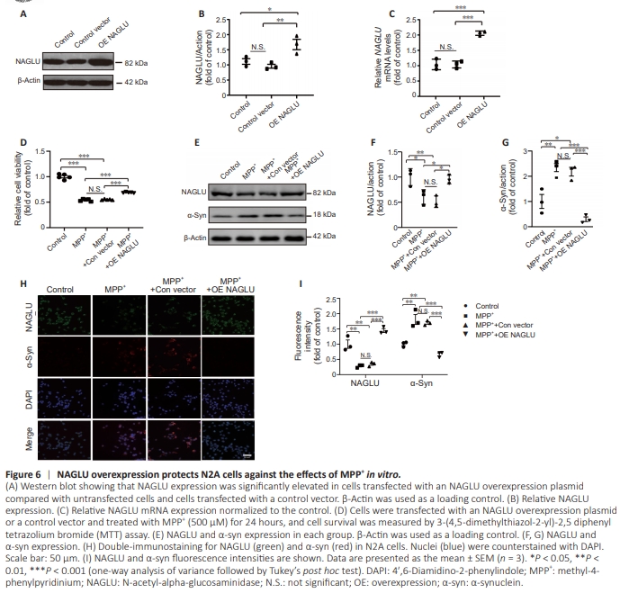
N-Acetyl-alpha-glucosaminidase overexpression alleviates 1-methyl-4-phenylpyridinium-induced cytotoxicity To further investigate the role of NAGLU in PD pathology, we overexpressed NAGLU in N2A cells and then treated them with MPP+ . As shown in Figure 6A–C, NAGLU protein and mRNA levels were significantly elevated in cells overexpressing NAGLU than in untransfected cells (P = 0.017) and cells transfected with a control vector (P = 0.006). MTT assay showed that MPP+ treatment significantly decreased the viability of N2A cells overexpressing NAGLU compared with untransfected cells (P < 0.001) and cells transfected with a control vector group (P < 0.001). Furthermore, NAGLU overexpression markedly improved the viability of N2A cells exposed to MPP+ -(compared with both untransfected N2A cells and cells transfected with a control vector) (P < 0.001; Figure 6D). N-Acetyl-alpha-glucosaminidase overexpression inhibits α-synuclein expression in 1-methyl-4-phenylpyridiniumtreated N2A cells WB analysis showed that α-syn expression was significantly downregulated, while NAGLU was significantly upregulated, in cells overexpressing NAGLU compared with cells transfected with a control vector after MPP+ treatment (P = 0.001; P = 0.014; Figure 6E–G). α-syn expression levels did not differ significantly between cells transfected with a control vector cells and untransfected cells exposed to MPP+ (P = 0.129; Figure 6E and G). Consistent with the WB results, the α-syn fluorescence intensity was significantly lower in MPP+ -treated cells overexpressing NAGLU than in MPP+ -treated cells transfected with a control vector (P < 0.001; Figure 6H and I). These findings indicate that NAGLU likely downregulates α-syn expression in PD.
Figure 7 | NAGLU expression was reduced in the brains of MPTP-treated mice.
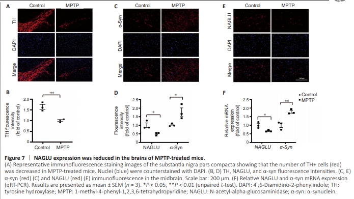
Consistent with a previous study (Zhang et al., 2022b), we found that tyrosine hydroxylase (TH) immunoreactivity was significantly reduced after MPTP treatment (P = 0.007; Figure 7A and B). Next, we performed immunofluorescence staining for NAGLU and α-syn in the MPTP and control groups. As depicted in Figure 7C and E, fewer cells in the substantia nigra exhibited NAGLU staining in the MPTP group than in the control group, indicating lower NAGLU expression in the MPTP group compared with the control group (P = 0.027). The MPTP group also showed a marked reduction in NAGLU mRNA expression (Figure 7F). In contrast, α-syn immunoreactivity (P = 0.038) and α-syn mRNA expression (P = 0.007) were significantly increased in the MPTP group (Figure 7C–F). These findings underscore the potential role of NAGLU in regulating α-syn in PD in vivo.