脊髓损伤
-
Figure 6 | Co-expression of the proliferation marker PCNA and GFP in oligodendrocyte clusters following SCI.
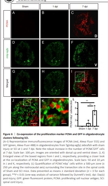
Next, we performed co-staining of PCNA, a proliferation marker, with green fluorescent protein (GPF) in Tg(mbp:egfp) zebrafish, in which mbp-expressing cells (oligodendrocyte cells) were labeled with EGFP (Chun et al., 2022). At 1 dpi, similar to sham zebrafish, no PCNA signal was detected (Figure 6A and B), despite the enrichment of cell proliferation pathways in single-cell RNAseq data at 1 dpi (Figure 5D). This discrepancy might reflect a lag between transcriptional- and protein-level changes. At 7 dpi, the significant increase in co-expression of the proliferation marker PCNA with mbp-driven GFP (mbp, myelin basic protein, a marker of oligodendrocyte) (Figure 6A–L) indicated that mature oligodendrocytes in Cluster 7 are actively proliferating.
Figure 7 | Proliferating oligodendrocytes upregulate CDH2 following SCI in zebrafish.
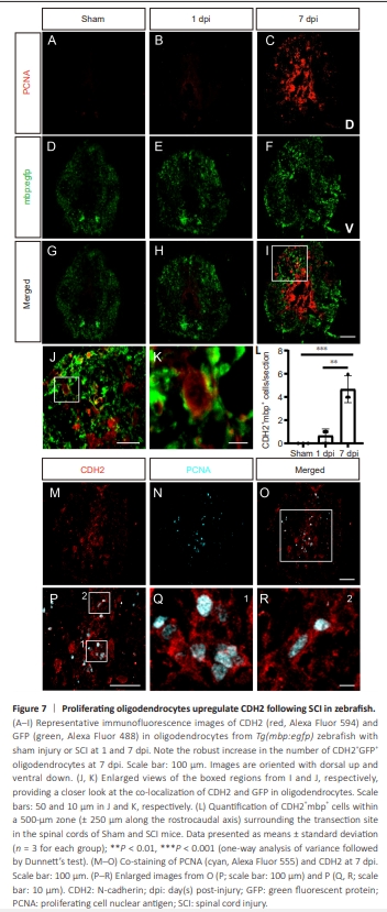
To confirm this activation of Cluster 7, we investigated the expression of cell adhesion molecule N-cadherin (CDH2), which is critical for stem cell activation during regeneration (Klatt Shaw et al., 2021). Indeed, CDH2 (cdh2) was found to be activated in Cluster 7 (Figure 5E). Furthermore, immunofluorescence staining revealed co-labeling of CDH2 with the oligodendrocyte marker MBP, showing a significant increase at 7 dpi (Figure 7A–L), similar to that observed for PCNA. CDH2 plays crucial roles in tissue morphogenesis, cell–cell adhesion, and maintenance of tissue architecture during development and regeneration (Klatt Shaw et al., 2021). The activation of CDH2 was confirmed by its coexpression with the proliferation marker PCNA (Figure 7M–R). Moreover, this dramatic post-SCI increase in CDH2 expression highlighted the activation and fate transition of oligodendrocytes.