神经退行性病
-
Figure 7 | Immunohistochemical analysis of microglia and astrocytes in the Hippocampus of male and female mice of NPC1mut mice.
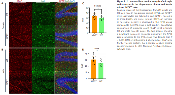
Next, we performed a similar analysis of tAge prediction on brain samples of APP/PS1 mouse model. Our analysis also showed accelerated biological tAge, increased mortality rate, and reduced relative expected lifespan (Figure 6 and Additional Figure 7). Similar trends in tAge observed in both models demonstrate accelerated aging and neuroinflammation, further bolstering the capability of NPC1mut model in recapitulating some aspects of AD-like phenotype. In addition, we subsequently analyzed the hippocampus of male and female NPC1mut mice and compared the microglial abundance in comparison to the WT controls (Figure 7A and B). Quantification of Iba1- positive cells (microglia) revealed a higher number of microglia in NPC1- mutant mice compared to control mice in both genders. Statistical analysis also showed greater significance in female mice (P = 0.029) compared to males (P = 0.039) further verifying the sex-specific effect of NPC1 mutation (Figure 7C and D). Overall, in the light of these commonalities between these two mouse models, our study thus represents a novel short-lived mouse model with accelerated brain aging to serve as an alternative mouse model for future application in fundamental and translational research of AD and brain aging.