脑损伤
-
Figure 2|Astrocytic ET-1 overexpression after MCAO promotes NSC proliferation in the DG region of the mouse hippocampus.
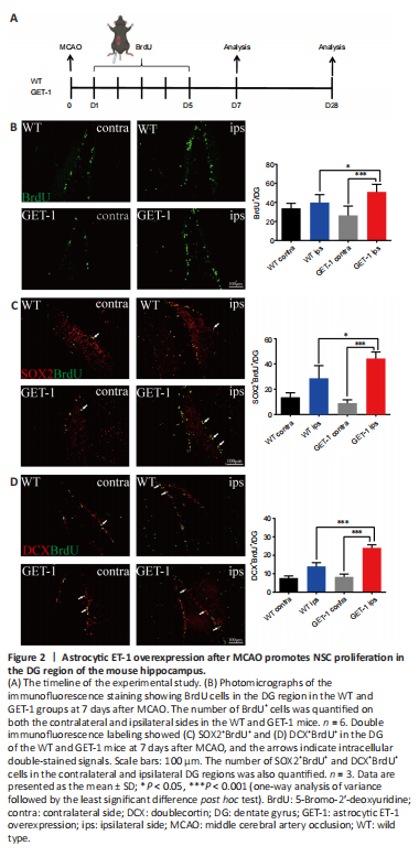
结果:Neurogenesis in the SGZ of the hippocampal DG is very important for cognitive function. We used BrdU as a measure of cell proliferation to confirm the influence of ET-1 overexpression on cell proliferation in both GET-1 and WT mice after ischemic stroke (Han et al., 2015; Bowers et al., 2020). We administered an intraperitoneal injection of BrdU to investigate DG cell proliferation at 7 days after ischemic stroke (Figure 2A). As shown in Figure 2B, in both the GET-1 and WT mice, the ipsilateral side (injured) expressed more BrdU+ cells than the contralateral side. Compared with the WT mice, the BrdU+ cells in the ipsilateral DG region of the GET-1 mice were significantly increased 7 days after ischemic injury (P = 0.026 for GET-1 ips vs. WT ips, P < 0.001 for GET-1 ips vs. GET-1 contra, Figure 2B). We conducted double-labeling with BrdU and SOX2 (a known phenotypic marker for NSCs) to identify the proliferating cells (Penisson et al., 2019). We found a conspicuous increase in SOX2+BrdU+ cells in the ipsilateral SGZ of GET-1 mice compared with the contralateral side 7 days after ischemic stroke. Ipsilateral SOX2+BrdU+ expression in the GET-1 mice was also higher than that in the WT mice (P = 0.013 for GET-1 ips vs. WT ips, P < 0.001 for GET-1 ips vs. GET-1 contra; Figure 2C). DCX is often used as a marker of migrating neuroblasts and early differentiated (immature) neurons (Brown et al., 2003). To further investigate whether overexpression of ET-1 in astrocytes was associated with neurogenesis in the DG region, including the regulation of neurogenesis, we performed BrdU and DCX double immunofluorescence labeling. At 7 days post-MCAO, there were more DCX+BrdU+ cells on the ipsilateral side than on the contralateral side in both GET-1 mice and WT mice, and this difference was significant in the GET-1 mice. Additionally, more DCX+BrdU+ cells were observed on the ipsilateral side in GET-1 mice than on the corresponding side in WT mice (P < 0.001 for GET-1 ips vs. WT ips, P < 0.001 for GET-1 ips vs. GET-1 contra; Figure 2D). Consequently, astrocytic ET-1 overexpression reinforced ischemia-induced NSC proliferation in the DG region of the hippocampus, as verified by our results.
Figure 3|Astrocytic ET-1 overexpression after MCAO impairs neurogenesis in the DG region.
结果:To determine whether NSCs in the WT and GET-1 mice differentiated into astrocytes or neurons after ischemic stroke, we conducted GFAP/BrdU and NeuN/BrdU costaining 28 days after MCAO. We found no significant differences between NeuN+BrdU+ and GFAP+BrdU+ costained cells on the contralateral (normal) side in GET-1 and WT mice (data not shown). However, at 28 days after MCAO, WT mice had more NeuN+BrdU+ cells in the ipsilateral DG region than GET-1 mice (P = 0.034 for GET-1 ips vs. WT ips; Figure 3A and C). In contrast, we observed slightly more GFAP+BrdU+ cells in GET-1 mice than in WT mice, but this difference was not significant (Figure 3B and D). This result suggests that ET-1 overexpression impairs neurogenesis in the DG region after cerebral ischemic injury (Figure 3E). In addition, Ki67 is a protein that labels cells that are actively dividing but not cells that have divided and have exited the cell cycle. We conducted Ki67/NeuN and Ki67/GFAP double immunofluorescence labeling at 3 months after ischemic stroke, and observed similar expression trends in Ki67+NeuN+ and Ki67+GFAP+ cells on the ipsilateral sides in WT and GET-1 mice (Additional Figure 2).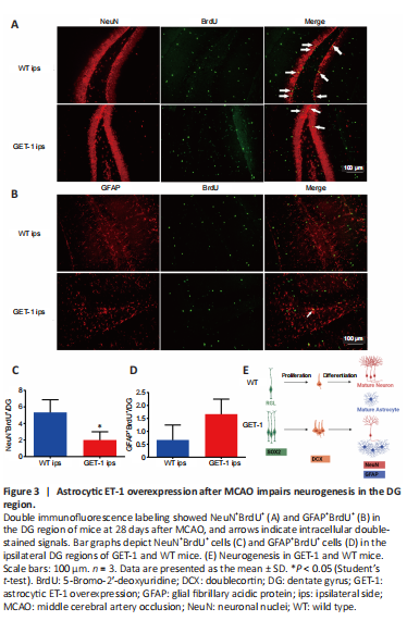
点击此处查看全文