神经损伤与修复
-
Figure 6|MT1R and MT2R are expressed on GABAergic neurons in Ctnnd2 KO mice.
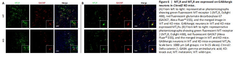
Figure 7|MT binds MT-Rs to improve GABAergic synaptic function in Ctnnd2 KO mice by activating PI3K/Akt signaling.
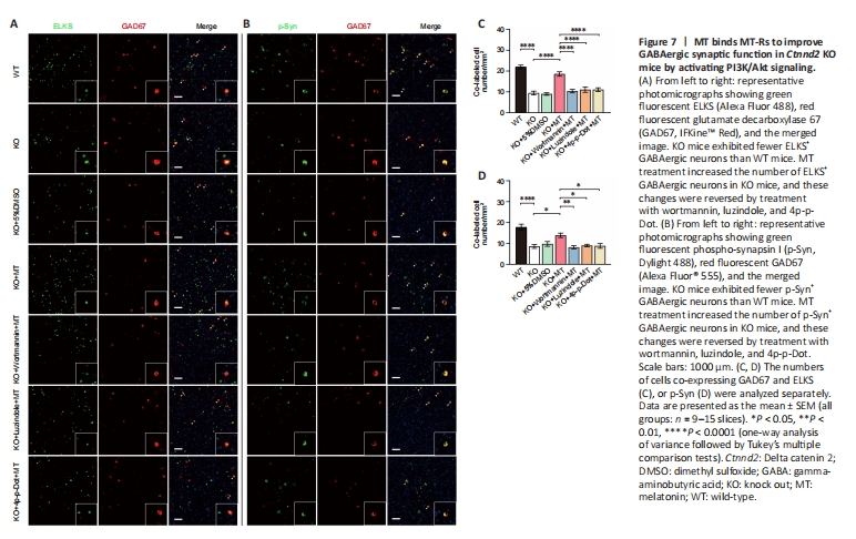
To investigate the effect of MT on GABAergic neurons in the PFC, we used immunofluorescence staining to examine MT1R, MT2R, and GAD67 co-expression. Our analysis indicated that GABAergic (GAD67-positive) neurons expressed both MT1Rs and MT2Rs (Figure 6A and B). Next, we asked whether MT promotes GABAergic neuron activity. Consistent with the western blot results, KO mice exhibited fewer ELKS+ (P < 0.0001) and p-Syn+ (P < 0.0001) GABAergic neurons than WT mice (Figure 7A–D). Local infusion with MT increased the numbers of ELKS+ and p-Syn+ GABAergic neurons, and these changes were reversed by treatment with wortmannin, luzindole, and 4p-p-Dot (ELKS: P < 0.0001 for KO vs. KO + MT group, P < 0.0001 for KO + wortmannin + MT vs. KO + MT group, P < 0.0001 for KO + luzindole + MT vs. KO + MT group, P < 0.0001 for KO + 4p-p-Dot + MT vs. KO + MT group; p-Syn: P = 0.0112 for KO vs. KO + MT group, P = 0.0032 for KO + wortmannin + MT vs. KO + MT group, P = 0.0319 for KO + luzindole + MT vs. KO + MT group, P = 0.0232 for KO + 4p-p-Dot + MT vs. KO + MT group, Figure 7A–D). Taken together, these results indicate that MT ameliorated GABAergic synaptic function by binding to MT-Rs on GABAergic neurons and activating the PI3K/Akt signaling pathway in Ctnnd2 KO mice.