脊髓损伤
-
Figure 2|BA ameliorates injury at the histopathological levels in the spinal cord of SCI rats.

H&E staining was performed to assessed damage at the histopathological levels in the spinal cords of SCI rats. Compared with the sham group, rats in the model group showed degeneration of spinal cord tissue neurons, damage to peripheral neurons, edema, degeneration of the myelin sheath in the white matter, and tissue structure disruption. BA and methylprednisolone improved all of these parameters (Figure 2A) and significantly reduced H&E staining scores in the spinal cord tissue of SCI rats compared with the findings in SCI rats (P < 0.01; Figure 2B). These findings indicate that BA ameliorates damage at the histopathological levels in the spinal rods of SCI rats.
Figure 3| On day 21 after injury, the effects of BA on neuronal apoptosis and neuron damage in SCI rats were observed.

TUNEL staining and Nissl staining were performed to observe the degree of apoptosis and damage of neurons in spinal cord tissue, respectively. As shown in Figure 3A and B, the green fluorescence signal was strongest in the model group. The TUNEL staining results showed that the apoptosis rate in the spinal cord tissue was higher in the model group than in the sham group; more importantly, the apoptosis rate decreased after treatment with BA or methylprednisolone (P < 0.01). The Nissl staining results showed that most of the neurons in the spinal cord tissue of rats in the model group were in a pyknotic hyperchromatic state; whereas, in rats treated with BA and methylprednisolone, the number of neurons with intact cell structure increased, and the number of Nissl bodies was significantly increased (P < 0.01; Figure 3C and D). These results indicate that BA treatment inhibits apoptosis and increases the number of intact neurons in SCI rats.
Figure 4|On day 21 after injury, the promoting effect of BA on neuronal autophagy in the spinal cord tissue of SCI rats was observed.
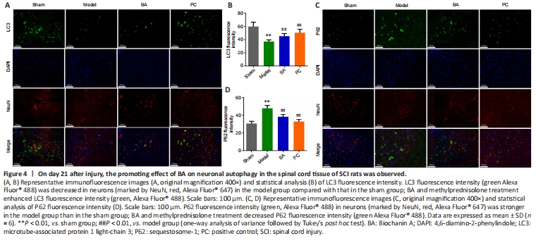
Next, immunofluorescence staining was used to measure LC3 and P62 expression in spinal cord tissue. Compared with that in the sham group, LC3 immunopositivity in the model group was significantly lower, while P62 immunopositivity was significantly higher (P < 0.01, Figure 4A–D). Interestingly, BA and methylprednisolone significantly increased LC3 immunopositivity and decreased P62 immunopositivity (P < 0.01, Figure 4A–D). These findings suggest that BA promotes neuronal autophagy in the spinal cord of SCI rats.
Figure 5|BA suppresses pyroptosis and promotes anti-oxidation in the spinal cord of SCI rats.
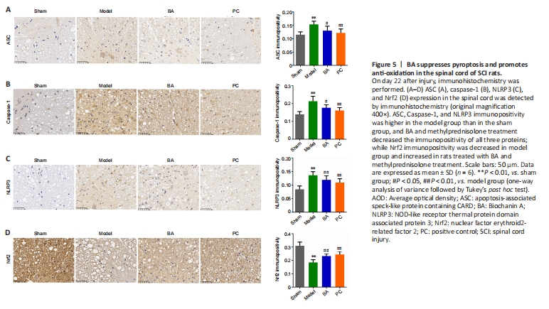
We next used immunohistochemistry to observe the expression levels of markers related to the inflammasome (ASC and NLRP3), pyroptosis (caspase-1), and antioxidation (Nrf2) in spinal cord tissue. The results showed that ASC, caspase-1, and NLRP3 immunopositivity was significantly higher, while Nrf2 expression was significantly lower, in the model group than in the sham group (all P < 0.01, Figure 5A–D). Importantly, ASC, caspase-1, and NLRP3 levels were lower in SCI rats than in sham rats, while BA and methylprednisolone treatment increased Nrf2 expression (all P < 0.01, Figure 5A–D). Moreover, qRT-PCR analysis demonstrated that the expression of genes related to the inflammasome (ASC and NLRP3) and pyroptosis (caspase-1 and GSDMD) in spinal cord tissue was higher in SCI rats compared with that in the sham group and decreased in SCI rats treated with BA and methylprednisolone (all P < 0.05, Figure 6A–D). These findings suggest that BA may reduce neuroinflammation and improve antioxidation in the spinal cord of SCI rats. Western blot analysis confirmed that BA or methylprednisolone effectively decreased the expression levels of the inflammasome-related proteins NLRP3, ASC, and GSDMD (all P < 0.05, Figure 6A–H), the expression levels of IL-18, IL-1β, and TLR4, and the ratios of p-P65/P65 and p-IκBα/IκBα in the spinal cord tissue of SCI rats compared with the findings in the model group (all P < 0.05, Figure 6E, I–M). These results show that BA suppresses pyroptosis, which may be related to the decreased inflammasome and increased antioxidation observed in the spinal cord of SCI rats.