中国神经再生研究(英文版) ›› 2023, Vol. 18 ›› Issue (11): 2406-2412.doi: 10.4103/1673-5374.371376

• 原著:脑损伤修复保护与再生 • 上一篇    下一篇

间充质干细胞来源细胞外囊泡修复创伤性中枢神经系统损伤:系统综述和meta分析


  • 出版日期:2023-11-15 发布日期:2023-05-04

Mesenchymal stem cell-derived extracellular vesicles therapy in traumatic central nervous system diseases: a systematic review and meta-analysis#br#

Zhelun Yang#, Zeyan Liang#, Jian Rao, Fabin Lin, Yike Lin, Xiongjie Xu, Chunhua Wang*, Chunmei Chen*   

  1. Department of Neurosurgery, Fujian Medical University Union Hospital, Fuzhou, Fujian Province, China
  • Online:2023-11-15 Published:2023-05-04
  • Contact: Chunmei Chen, MD, 1731012948@qq.com; Chunhua Wang, MD, wchmail@126.com.

摘要:

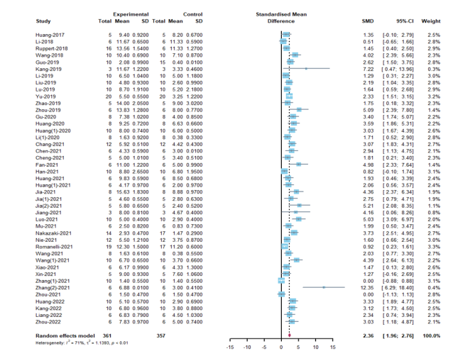
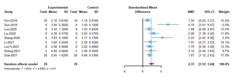
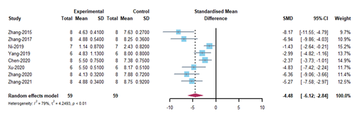
间充质干细胞来源的细胞外囊泡在治疗创伤性中枢神经系统损伤方面已被证明是一种有前途的非细胞疗法。然而,临床前的研究成果要向临床转化需要全方位和系统地掌握相关实验方法和了解间充质干细胞来源的细胞外囊泡对创伤性中枢神经系统损伤的疗效。因此,对近期使用间充质干细胞来源的细胞外囊泡治疗创伤性中枢神经系统损伤的动物试验进行了系统综述和meta分析。通过检索PubMed,Web of Science,The Cochrane Library和Ovid-Embase数据库截止2022年4月1日的数据,筛选间充质干细胞来源的细胞外囊泡治疗创伤性中枢神经系统疾病的临床前研究。应用SYRCLE的偏倚风险工具分析动物研究中的发表偏倚风险。最终60项研究被纳入研究,其中脊髓损伤研究52项,创伤性脑损伤研究8项。结果显示,与对照组相比,间充质干细胞来源的细胞外囊泡治疗可明显促进SCI动物的运动功能恢复,包括大鼠Basso Beattie Bresnahan评分(SMD: 2.36, 95% CI: 1.96-2.76, I² = 71%, P < 0.01)和小鼠BMS评分(SMD: 2.31, 95% CI: 1.57 - 3.04, I² = 60%, P = 0.01);可明显促进创伤性脑损伤动物神经功能的恢复,包括mNSS(SMD: - 4.48, 95% CI: - 6.12 to - 2.84, P < 0.01, I² = 79%)和Foot Fault Test(SMD: - 3.26, 95% CI: - 4.09 to - 2.42, I² = 21%, P = 0.28)。亚组分析显示,对于BBB评分而言,同种异体间充质干细胞来源的细胞外囊泡的疗效高于异种异体间充质干细胞来源的细胞外囊泡(同种异体: SMD: 2.54, 95% CI: 2.05 - 3.02, I² = 65.5%; 异种异体: SMD: 1.78, 95% CI: 1.1 - 2.45, I² = 74.6%; P = 0.0116);通过超滤离心联合密度梯度超速离心分离的间充质干细胞来源的细胞外囊泡(SMD:3.58,95%CI:2.62 - 4.53,I² = 31%,P < 0.0001)可能更有效。对于BMS评分而言,胎盘间充质干细胞来源的细胞外囊泡比骨髓来源的效果更好(胎盘:SMD: 5.25,95%CI:2.45 - 8.06,I² = 0%;骨髓:SMD: 1.82, 95% CI: 1.23 - 2.41, I² = 0%; P = 0.0421)。对于mNSS评分而言,骨髓间充质干细胞来源的细胞外囊泡比脂肪来源的效果更好(骨髓:SMD:-4.86,95%CI:-6.66-3.06,I²=81%;脂肪:SMD:-2.37,95%CI:-3.73-1.01,I²=0%;P=0.0306)。静脉给药(SMD: -5.47, 95% CI: -6.98 - -3.97, I² = 53.3%, P = 0.0002)和给药剂量等于100μg(SMD: -5.47, 95% CI: -6.98 - -3.97, I² = 53.3%, P < 0.0001)显示出更好的结果。各项研究的异质性很小,敏感性分析也表明结果稳定。最后,所有实验的方法学质量大多令人满意。总之,间充质干细胞来源的细胞外囊泡在促进创伤性中枢神经系统损伤后运动和神经功能恢复方面发挥了关键作用。

https://orcid.org/0000-0002-4490-0380 (Chunmei Chen); https://orcid.org/0000-0002-4483-7465 (Chunhua Wang)

Abstract: Although there are challenges in treating traumatic central nervous system diseases, mesenchymal stem cell-derived extracellular vesicles (MSC-EVs) have recently proven to be a promising non-cellular therapy. We comprehensively evaluated the efficacy of mesenchymal stem cell-derived extracellular vesicles in traumatic central nervous system diseases in this meta-analysis based on preclinical studies. Our meta-analysis was registered at PROSPERO (CRD42022327904, May 24, 2022). To fully retrieve the most relevant articles, the following databases were thoroughly searched: PubMed, Web of Science, The Cochrane Library, and Ovid-Embase (up to April 1, 2022). The included studies were preclinical studies of mesenchymal stem cell-derived extracellular vesicles for traumatic central nervous system diseases. The Systematic Review Centre for Laboratory Animal Experimentation (SYRCLE)’s risk of bias tool was used to examine the risk of publication bias in animal studies. After screening 2347 studies, 60 studies were included in this study. A meta‐analysis was conducted for spinal cord injury (n = 52) and traumatic brain injury (n = 8). The results indicated that mesenchymal stem cell-derived extracellular vesicles treatment prominently promoted motor function recovery in spinal cord injury animals, including rat Basso, Beattie and Bresnahan locomotor rating scale scores (standardized mean difference [SMD]: 2.36, 95% confidence interval [CI]: 1.96–2.76, P < 0.01, I2 = 71%) and mouse Basso Mouse Scale scores (SMD = 2.31, 95% CI: 1.57–3.04, P = 0.01, I2 = 60%) compared with controls. Further, mesenchymal stem cell-derived extracellular vesicles treatment significantly promoted neurological recovery in traumatic brain injury animals, including the modified Neurological Severity Score (SMD = –4.48, 95% CI: –6.12 to –2.84, P < 0.01, I2 = 79%) and Foot Fault Test (SMD = –3.26, 95% CI: –4.09 to –2.42, P = 0.28, I2 = 21%) compared with controls. Subgroup analyses showed that characteristics may be related to the therapeutic effect of mesenchymal stem cell-derived extracellular vesicles. For Basso, Beattie and Bresnahan locomotor rating scale scores, the efficacy of allogeneic mesenchymal stem cell-derived extracellular vesicles was higher than that of xenogeneic mesenchymal stem cell-derived extracellular vesicles (allogeneic: SMD = 2.54, 95% CI: 2.05–3.02, P = 0.0116, I2 = 65.5%; xenogeneic: SMD: 1.78, 95%CI: 1.1–2.45, P = 0.0116, I2 = 74.6%). Mesenchymal stem cell-derived extracellular vesicles separated by ultrafiltration centrifugation combined with density gradient ultracentrifugation (SMD = 3.58, 95% CI: 2.62–4.53, P < 0.0001, I2 = 31%) may be more effective than other EV isolation methods. For mouse Basso Mouse Scale scores, placenta-derived mesenchymal stem cell-derived extracellular vesicles worked better than bone mesenchymal stem cell-derived extracellular vesicles (placenta: SMD = 5.25, 95% CI: 2.45–8.06, P = 0.0421, I2 = 0%; bone marrow: SMD = 1.82, 95% CI: 1.23–2.41, P = 0.0421, I2 = 0%). For modified Neurological Severity Score, bone marrow-derived MSC-EVs worked better than adipose-derived MSC-EVs (bone marrow: SMD = –4.86, 95% CI: –6.66 to –3.06, P = 0.0306, I2 = 81%; adipose: SMD = –2.37, 95% CI: –3.73 to –1.01, P = 0.0306, I2 = 0%). Intravenous administration (SMD = –5.47, 95% CI: –6.98 to –3.97, P = 0.0002, I2 = 53.3%) and dose of administration equal to 100 μg (SMD = –5.47, 95% CI: –6.98 to –3.97, P < 0.0001, I2 = 53.3%) showed better results than other administration routes and doses. The heterogeneity of studies was small, and sensitivity analysis also indicated stable results. Last, the methodological quality of all trials was mostly satisfactory. In conclusion, in the treatment of traumatic central nervous system diseases, mesenchymal stem cell-derived extracellular vesicles may play a crucial role in promoting motor function recovery.

Key words: animals, central nervous system diseases, extracellular vesicles, mesenchymal stromal cell, meta-analysis, spinal cord injury, traumatic brain injury