中国神经再生研究(英文版) ›› 2024, Vol. 19 ›› Issue (1): 220-225.doi: 10.4103/1673-5374.375341

• 原著:视神经损伤修复保护与再生 • 上一篇    下一篇

青光眼模型初级视觉皮质神经元的形态特征及视觉调谐功能


  • 出版日期:2024-01-15 发布日期:2023-08-02

Morphological disruption and visual tuning alterations in the primary visual cortex in glaucoma (DBA/2J) mice

Yin Yang1, #, Zhaoxi Yang1, #, Maoxia Lv1, #, Ang Jia2, Junjun Li2, Baitao Liao2, Jing’an Chen3, 4, Zhengzheng Wu1, Yi Shi5, 6, Yang Xia1, 2, Dezhong Yao1, 2, 3, 7, *, Ke Chen1, 2, *   

  1. 1Department of Ophthalmology, Sichuan Provincial People’s Hospital, Medical School, University of Electronic Science and Technology of China, Chengdu, Sichuan Province, China; 2The Clinical Hospital of Chengdu Brain Science Institute, MOE Key Lab for NeuroInformation, School of Life Science and Technology, University of Electronic Science and Technology of China, Chengdu, Sichuan Province, China; 3Research Unit of NeuroInformation, Chinese Academy of Medical Sciences, Chengdu, Sichuan Province, China; 4Institute of Basic Medical Sciences Chinese Academy of Medical Sciences, School of Basic Medicine Peking Union Medical College, Beijing, China; 5Health Management Center, Sichuan Provincial Key Laboratory for Human Disease Gene Study, Sichuan Provincial People’s Hospital, University of Electronic Science and Technology of China, Chengdu, Sichuan Province, China; 6Research Unit for Blindness Prevention of Chinese Academy of Medical Sciences (2019RU026), Sichuan Academy of Medical Sciences, Chengdu, Sichuan Province, China; 7School of Electrical Engineering, Zhengzhou University, Zhengzhou, Henan Province, China
  • Online:2024-01-15 Published:2023-08-02
  • Contact: Dezhong Yao, PhD, dyao@uestc.edu.cn; Ke Chen, PhD, chenke0703@uestc.edu.cn.
  • Supported by:
    This study was supported by the STI 2030–Major Projects 2022ZD0208500 (to DY); the National Natural Science Foundation of China, Nos. 82072011 (to YX), 82121003 (to DY), 82271120 (to YS); Sichuan Science and Technology Program, No. 2022ZYD0066 (to YS); a grant from Chinese Academy of Medical Science, No. 2019-12M-5-032 (to YS); and the Fundamental Research Funds for the Central Universities, No. ZYGX2021YGLH219 (to KC).

摘要:

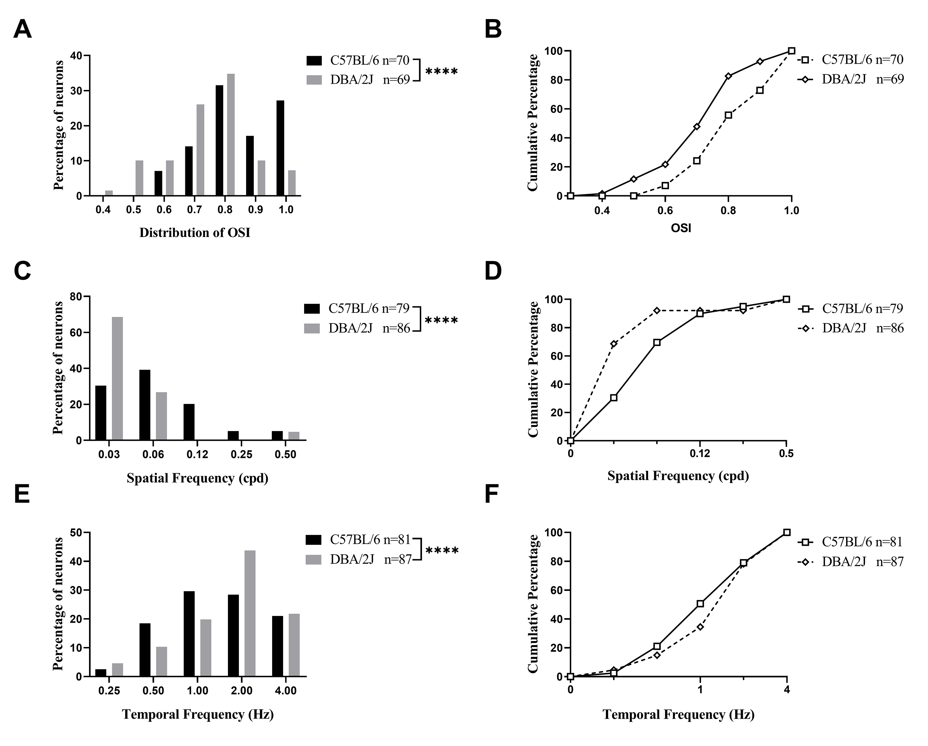
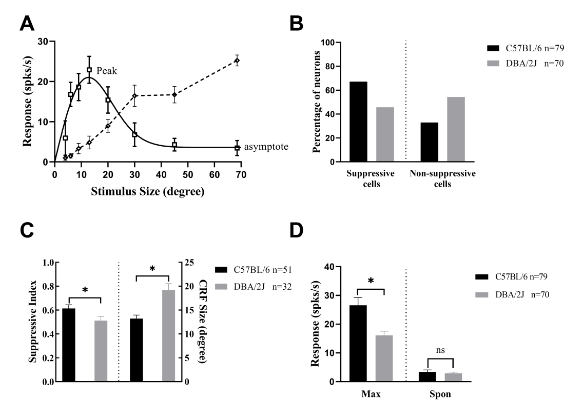
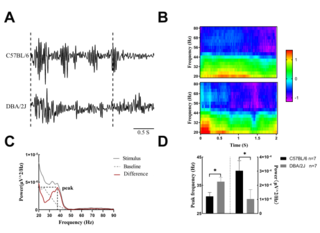
随着青光眼研究的深入,越来越多的研究关注到青光眼患者的视觉皮质甚至整个大脑的变化。但很少有研究涉及在青光眼模型中眼部退化导致大脑的结构与功能的改变 。因此,在青光眼模型中对初级视觉皮质神经元的形态特征及视觉调谐功能进行探究,可以增加对“脑器(眼)交互”中眼睛退化与大脑损伤的联系及病理机制的理解。实验以DBA/2J小鼠作为自发性继发性青光眼模型。通过对青光眼小鼠(DBA/2J)和年龄匹配的正常小鼠(C57BL/6J)初级视觉皮质神经元的组织形态学和电生理学响应特征的比较研究。小鼠V1脑片NeuN 染色和 Nissl染色结果显示,青光眼小鼠V1中观察到的神经元数量显著减少,神经元内尼氏小体密度降低。首先对V1神经元的视觉调谐曲线测试的结果发现,与正常小鼠相比,青光眼小鼠的视觉调谐特征出现了损伤:方位选择性减弱、偏好低的空间频率和高时间频率的刺激光栅,此外通过空间整合测试发现大部分神经元的外周抑制作用的强度更低、而神经元的感受野的范围增大,而视觉刺激诱导的伽马节律振荡能量也会显著减弱。该研究通过直接测试初级视觉皮质区细胞的电生理反应属性和解析其形态结构,丰富了“眼损伤对大脑影响”这个问题理解,从而为青光眼的治疗提供新视角。

https://orcid.org/0000-0002-8042-879X (Dezhong Yao); https://orcid.org/0000-0003-2209-1452 (Ke Chen)

Abstract: Glaucoma is a leading cause of irreversible blindness worldwide, and previous studies have shown that, in addition to affecting the eyes, it also causes abnormalities in the brain. However, it is not yet clear how the primary visual cortex (V1) is altered in glaucoma. This study used DBA/2J mice as a model for spontaneous secondary glaucoma. The aim of the study was to compare the electrophysiological and histomorphological characteristics of neurons in the V1 between 9-month-old DBA/2J mice and age-matched C57BL/6J mice. We conducted single-unit recordings in the V1 of light-anesthetized mice to measure the visually induced responses, including single-unit spiking and gamma band oscillations. The morphology of layer II/III neurons was determined by neuronal nuclear antigen staining and Nissl staining of brain tissue sections. Eighty-seven neurons from eight DBA/2J mice and eighty-one neurons from eight C57BL/6J mice were examined. Compared with the C57BL/6J group, V1 neurons in the DBA/2J group exhibited weaker visual tuning and impaired spatial summation. Moreover, fewer neurons were observed in the V1 of DBA/2J mice compared with C57BL/6J mice. These findings suggest that DBA/2J mice have fewer neurons in the V1 compared with C57BL/6J mice, and that these neurons have impaired visual tuning. Our findings provide a better understanding of the pathological changes that occur in V1 neuron function and morphology in the DBA/2J mouse model. This study might offer some innovative perspectives regarding the treatment of glaucoma. 

Key words: DBA/2J, degeneration, gamma band oscillations, glaucoma, primary visual cortex (V1), retina, single-unit recording, tuning curve