中国神经再生研究(英文版) ›› 2026, Vol. 21 ›› Issue (9): 4352-4357.doi: 10.4103/NRR.NRR-D-25-00685

• 原著:脑损伤修复保护与再生 • 上一篇    下一篇

布洛卡区负责言语生成功能受肺功能调节


  • 出版日期:2026-09-15 发布日期:2026-05-22

Broca’s area, responsible for speech production, is regulated by lung function

Siyu Cao1, #, Wenwen Zhuang2, 3, #, Yuqian Hu3, #, Shijun Qiu4, *, Li-Hai Tan2, 3, 5, 6, *   

  1. 1Department of Respiratory and Critical Care Medicine, Peking University Third Hospital, Beijing, China; 
    2Guangdong-Hongkong-Macau Institute of CNS Regeneration and Key Laboratory of CNS Regeneration (Ministry of Education), Jinan University, Shenzhen Campus, Shenzhen, Guangdong Province, China; 
    3Center for Language and Brain, Shenzhen Institute of Neuroscience, Shenzhen, Guangdong Province, China; 
    4Department of Radiology, First Affiliated Hospital of Guangzhou University of Chinese Medicine, Guangzhou, Guangdong Province, China; 
    5Guangdong Innovation Platform of Translational Research for Cerebrovascular Diseases, Shenzhen, Guangdong Province, China; 
    6University International College, Macau University of Science and Technology, Macao Special Administrative Region, China
  • Online:2026-09-15 Published:2026-05-22
  • Contact: Li-Hai Tan, PhD, tanlh@sions.cn; Shijun Qiu, MD, qiu-sj@163.com.
  • Supported by:
    This study was supported by the Brain Disorders and Protection of Cortical Language Functions, No. 2016ZT06S220 (to LHT).

摘要:

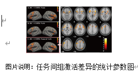
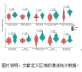
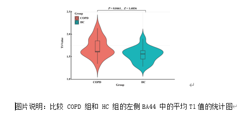
人们已知布洛卡区(特别是左下额回的盖部与三角部)对人类言语生成至关重要。然而该区域为何参与言语活动仍未可知。言语生成不仅涉及概念构思与运动规划,还需依靠呼吸系统提供发声所需的气流。因此,布洛卡区在言语中的作用可能受肺部功能及相关脑区调控。为验证此假设,试验招募慢性阻塞性肺病(COPD)患者,在功能性磁共振成像(fMRI)与定量磁共振成像(qMRI)扫描过程中要求其进行言语任务。结果观察到,COPD患者在执行言语任务时,左侧下额叶皮质及其他区域的皮质反应发生改变,与呼吸相关的皮质区域出现异常激活。此外,通过qMRI生成长程弛豫时间(T1)图谱作为脑微结构变化指标(包括树突成熟与髓鞘化),发现患者布洛卡区T1值显著高于对照组,表明该区域髓鞘化程度降低且微结构完整性受损。关键数据表明:呼吸困难程度越严重,布洛卡区微结构发育越不完善且激活强度越弱。此研究首次证实肺部功能可塑造布洛卡区作为言语中枢的特性,为肺功能障碍影响皮层语言区功能与结构特性提供了新证据。这些发现凸显了肺脑轴在言语生成中的机制作用,并为改善言语表现的干预措施提供了潜在靶点。


https://orcid.org/0000-0001-6983-1767 (Li-Hai Tan); 

https://orcid.org/0000-0002-6739-4265 (Shijun Qiu)

关键词: 布洛卡区l言语生成l肺功能l功能磁共振成像l定量磁共振成像l慢性阻塞性肺病l肺脑轴l呼吸困难l微结构改变lT1

Abstract: For more than 150 years, Broca’s area—specifically, the pars opercularis and pars triangularis in the left inferior frontal gyrus—has been recognized as crucial for human speech production. However, it remains unknown why this region is recruited for speaking. Speech production involves not only conceptualization and motor planning but also respiration to provide the necessary airflow for creating sounds. Thus, the role of Broca’s area in speech may be shaped by the functionality and related brain regions of the lungs. To test this hypothesis, we recruited patients with chronic obstructive pulmonary disease and asked them to read words aloud while their brains were scanned using functional magnetic resonance imaging, with quantitative magnetic resonance imaging acquired separately. The chronic obstructive pulmonary disease patients exhibited altered cortical responses in the left inferior prefrontal cortex and other regions during speech tasks, and also had abnormal activation in cortical sites associated with breathing. In addition, using quantitative magnetic resonance imaging to generate longitudinal relaxation time (T1) maps as an index of brain microstructural changes, including dendritic maturation and myelination, we observed significantly longer longitudinal relaxation times in Broca’s area in the chronic obstructive pulmonary disease group than in the control group, suggesting reduced myelination and impaired microstructural integrity. Crucially, our data indicated that more severe dyspnea was associated with less well-developed microstructure in Broca’s area and weaker activation of this region. The present study indicates for the first time that the lungs may function to shape Broca’s area as the speaking center, providing novel evidence that pulmonary dysfunction can influence both the functional and structural properties of cortical language regions. These findings highlight the mechanistic role of the lung–brain axis in speech production and suggest potential targets for interventions to enhance speech performance.

Key words: Broca’s areal chronic obstructive pulmonary diseasel dyspneal functional magnetic resonance imagingl lung-brain axisl speech productionl lung functionsl microstructural changesl quantitative magnetic resonance imaging