中国神经再生研究(英文版) ›› 2025, Vol. 20 ›› Issue (5): 1495-1504.doi: 10.4103/NRR.NRR-D-23-00493

• 原著:周围神经损伤修复保护与再生 • 上一篇    下一篇

不同神经重建策略下静息态脑网络重塑:臂丛神经损伤大鼠功能磁共振成像分析


  • 出版日期:2025-05-15 发布日期:2024-10-31
  • 基金资助:
    国家自然科学基金项目81871836,82172554和81802249,81902301;国家重点研发计划项目2018YFC2001600和2018YFC2001604;上海市新星计划项目19QA1409000;上海市卫生和计划生育委员会项目2018YQ02;上海市青年拔尖人才培养计划和上海市 “新星计划 ”资助。19QA1409000;上海市卫生和计划生育委员会2018YQ02;以及上海市青年拔尖人才发展计划和上海市 “医学英才新星 ”青年发展计划RY411.19.01.10

Resting-state brain network remodeling after different nerve reconstruction surgeries: a functional magnetic resonance imaging study in brachial plexus injury rats

Yunttng Xiang1, 2, # , Xiangxin Xing3, # , Xuyun Hua3, 4, # , Yuwen Zhang5 , Xin Xue1 , Jiajia Wu3, 4 , Mouxiong Zheng3, 4, * , He Wang5, 6, 7, * , Jianguang Xu1, 2, 3, *   

  1. 1 School of Rehabilitatton Science, Shanghai University of Tradittonal Chinese Medicine, Shanghai, China;  2 Engineering Research Center of Tradittonal Chinese Medicine Intelligent Rehabilitatton, Ministry of Educatton, Shanghai, China;  3 Department of Rehabilitatton Medicine, Yueyang Hospital of Integrated Tradittonal Chinese and Western Medicine, Shanghai University of Tradittonal Chinese Medicine, Shanghai, China;  4 Department of Traumatology and Orthopedics, Yueyang Hospital of Integrated Tradittonal Chinese and Western Medicine, Shanghai University of Tradittonal Chinese Medicine, Shanghai, China;  5 Instttute of Science and Technology for Brain-Inspired Intelligence, Fudan University, Shanghai, China;  6 Key Laboratory of Computattonal Neuroscience and Brain-Inspired Intelligence (Fudan University), Ministry of Educatton, Shanghai, China; 7 Human Phenome Instttute, Fudan University, Shanghai, China
  • Online:2025-05-15 Published:2024-10-31
  • Contact: Jianguang Xu, MD, PhD, xjg@shutcm.edu.cn; He Wang, PhD, hewang@fudan.edu.cn; Mouxiong Zheng, MD, PhD, zhengmouxiong@shutcm.edu.cn.
  • Supported by:
    This work was supported by the Nattonal Natural Science Foundatton of China, Nos. 81871836 (to MZ), 82172554 (to XH), and 81802249 (to XH), 81902301 (to JW); the Nattonal Key R&D Program of China, Nos. 2018YFC2001600 (to JX) and 2018YFC2001604 (to JX); Shanghai Rising Star Program, No. 19QA1409000 (to MZ); Shanghai Municipal Commission of Health and Family Planning, No. 2018YQ02 (to MZ); and Shanghai Youth Top Talent Development Plan and Shanghai “Rising Stars of Medical Talent” Youth Development Program, No. RY411.19.01.10 (to XH).

摘要:

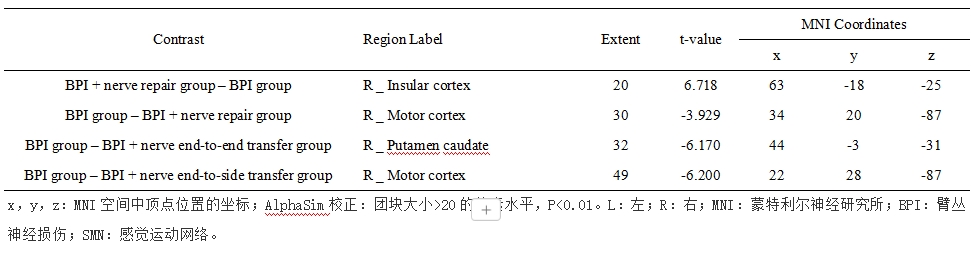
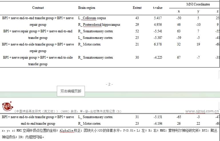
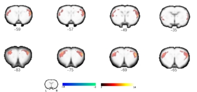
既往研究显示,传统的神经端到端移位后,大脑控制患肢运动的皮质区逐渐从供体神经转移到受体神经,涉及复杂的大脑重塑过程。而神经端到侧移位可以在不牺牲供体神经的情况下恢复受体神经功能。目前对于神经重建后患肢运动表征的过程和在此过程中的关键脑区都得到研究的证实,但不同神经重建策略在大脑网络水平的异同尚未阐明。实验给予Sprague-Dawley大鼠左臂丛神经完全切断后,进行以下干预:无神经修复、移植神经修复、膈神经末端转移和末端侧转移并将移植神经缝合到上干前部。术后 7 个月采集静息态脑功能磁共振图像。利用独立成分分析算法确定感兴趣的组级网络成分,并提取成分内每个体素的静息态功能连通性(RS FC)值。比较四组之间网络内 RS FC 的变化。通过行为观察(肘关节屈曲)和肌电图评估靶肌肉再支配。实验发现感觉运动网络和内感觉网络是周围神经通路重建后大脑重塑的关键功能网络,不同神经重建术之间大脑功能网络的改变存在不同的中枢机制。神经修复与感觉运动网络的内在连接性增强有关,而神经端到侧转移可能更有利于恢复原有运动表象对患肢的控制。神经修复和神经端到端转移后,丘脑-皮质通路在内感觉网络内增强,而神经端到侧转移后,与认知和情感相关的脑区增强。该研究揭示了与周围神经损伤后不同神经重建相关的重要大脑网络,这些网络可能是进一步运动恢复的潜在调节目标。

https://orcid.org/0000-0002-9861-6086 (Jianguang Xu); https://orcid.org/0000-0002-2053-9439 (He Wang); https://orcid.org/0000-0003-0601-3403 (Mouxiong Zheng)

Abstract:

Distinct brain remodeling has been found after different nerve reconstruction strategies, including motor representation of the affected limb. However, differences among reconstruction strategies at the brain network level have not been elucidated. This study aimed to explore intra-network changes related to altered peripheral neural pathways after different nerve reconstruction surgeries, including nerve repair, end-to-end nerve transfer, and end-to-side nerve transfer. Sprague–Dawley rats underwent complete left brachial plexus transection and were divided into four equal groups of eight: no nerve repair, grafted nerve repair, phrenic nerve end-to-end transfer, and end-to-side transfer with a graft sutured to the anterior upper trunk. Resting-state brain functional magnetic resonance imaging was obtained 7 months after surgery. The independent component analysis algorithm was utilized to identify group-level network components of interest and extract resting-state functional connectivity values of each voxel within the component. Alterations in intra-network resting-state functional connectivity were compared among the groups. Target muscle reinnervation was assessed by behavioral observation (elbow flexion) and electromyography. The results showed that alterations in the sensorimotor and interoception networks were mostly related to changes in the peripheral neural pathway. Nerve repair was related to enhanced connectivity within the sensorimotor network, while end-to-side nerve transfer might be more beneficial for restoring control over the affected limb by the original motor representation. The thalamic-cortical pathway was enhanced within the interoception network after nerve repair and end-to-end nerve transfer. Brain areas related to cognition and emotion were enhanced after end-to-side nerve transfer. Our study revealed important brain networks related to different nerve reconstructions. These networks may be potential targets for enhancing motor recovery. 

Key words:

brain functional networks, end-to-end nerve transfer, end-to-side nerve transfer, independent component analysis, nerve repair,  , peripheral plexus injury, resting-state functional connectivity